2SRC

CRYSTAL STRUCTURE OF HUMAN TYROSINE-PROTEIN KINASE C-SRC, IN COMPLEX WITH AMP-PNP


Experimental Data Snapshot

  • Method: X-RAY DIFFRACTION
  • Resolution: 1.50 Å
  • R-Value Free: 0.281 
  • R-Value Work: 0.226 
  • R-Value Observed: 0.226 

wwPDB Validation   3D Report Full Report


This is version 1.3 of the entry. See complete history


Literature

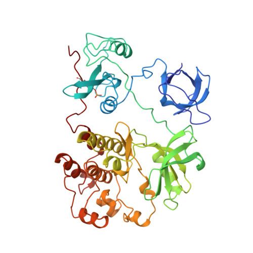
Crystal structures of c-Src reveal features of its autoinhibitory mechanism.

Xu, W.Doshi, A.Lei, M.Eck, M.J.Harrison, S.C.

(1999) Mol Cell 3: 629-638

  • DOI: https://doi.org/10.1016/s1097-2765(00)80356-1
  • Primary Citation of Related Structures:  
    2SRC

  • PubMed Abstract: 

    Src family kinases are maintained in an assembled, inactive conformation by intramolecular interactions of their SH2 and SH3 domains. Full catalytic activity requires release of these restraints as well as phosphorylation of Tyr-416 in the activation loop. In previous structures of inactive Src kinases, Tyr-416 and flanking residues are disordered. We report here four additional c-Src structures in which this segment adopts an ordered but inhibitory conformation. The ordered activation loop forms an alpha helix that stabilizes the inactive conformation of the kinase domain, blocks the peptide substrate-binding site, and prevents Tyr-416 phosphorylation. Disassembly of the regulatory domains, induced by SH2 or SH3 ligands, or by dephosphorylation of Tyr-527, could lead to exposure and phosphorylation of Tyr-416.


  • Organizational Affiliation

    Laboratory of Molecular Medicine, Children's Hospital, Boston, Massachusetts 02115, USA.


Macromolecules
Find similar proteins by:  (by identity cutoff)  |  3D Structure
Entity ID: 1
MoleculeChains Sequence LengthOrganismDetailsImage
TYROSINE-PROTEIN KINASE SRC452Homo sapiensMutation(s): 1 
EC: 2.7.1.112
UniProt & NIH Common Fund Data Resources
Find proteins for P12931 (Homo sapiens)
Explore P12931 
Go to UniProtKB:  P12931
PHAROS:  P12931
GTEx:  ENSG00000197122 
Entity Groups  
Sequence Clusters30% Identity50% Identity70% Identity90% Identity95% Identity100% Identity
UniProt GroupP12931
Sequence Annotations
Expand
  • Reference Sequence
Small Molecules
Ligands 1 Unique
IDChains Name / Formula / InChI Key2D Diagram3D Interactions
ANP
Query on ANP

Download Ideal Coordinates CCD File 
B [auth A]PHOSPHOAMINOPHOSPHONIC ACID-ADENYLATE ESTER
C10 H17 N6 O12 P3
PVKSNHVPLWYQGJ-KQYNXXCUSA-N
Modified Residues  1 Unique
IDChains TypeFormula2D DiagramParent
PTR
Query on PTR
A
L-PEPTIDE LINKINGC9 H12 N O6 PTYR
Binding Affinity Annotations 
IDSourceBinding Affinity
PTR BindingDB:  2SRC -TΔS: -1.81e+1 (kJ/mol) from 1 assay(s)
ΔG: -2.14e+1 (kJ/mol) from 1 assay(s)
Experimental Data & Validation

Experimental Data

  • Method: X-RAY DIFFRACTION
  • Resolution: 1.50 Å
  • R-Value Free: 0.281 
  • R-Value Work: 0.226 
  • R-Value Observed: 0.226 
  • Space Group: P 21 21 21
Unit Cell:
Length ( Å )Angle ( ˚ )
a = 50.59α = 90
b = 72.97β = 90
c = 172.69γ = 90
Software Package:
Software NamePurpose
X-PLORmodel building
X-PLORrefinement
X-PLORphasing

Structure Validation

View Full Validation Report


Entry History 

Deposition Data

Revision History  (Full details and data files)

  • Version 1.0: 1999-07-22
    Type: Initial release
  • Version 1.1: 2008-03-25
    Changes: Version format compliance
  • Version 1.2: 2011-07-13
    Changes: Version format compliance
  • Version 1.3: 2023-08-09
    Changes: Database references, Derived calculations, Other, Refinement description