1F0R

CRYSTAL STRUCTURE OF HUMAN COAGULATION FACTOR XA COMPLEXED WITH RPR208815


Experimental Data Snapshot

  • Method: X-RAY DIFFRACTION
  • Resolution: 2.10 Å
  • R-Value Free: 0.263 
  • R-Value Work: 0.216 
  • R-Value Observed: 0.216 

wwPDB Validation   3D Report Full Report


Ligand Structure Quality Assessment 


This is version 1.3 of the entry. See complete history


Literature

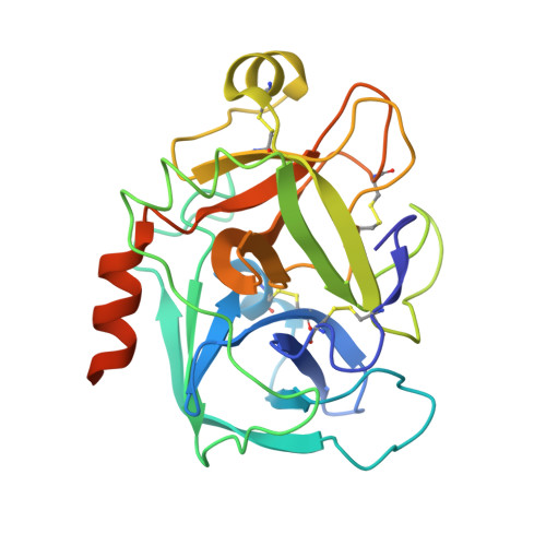
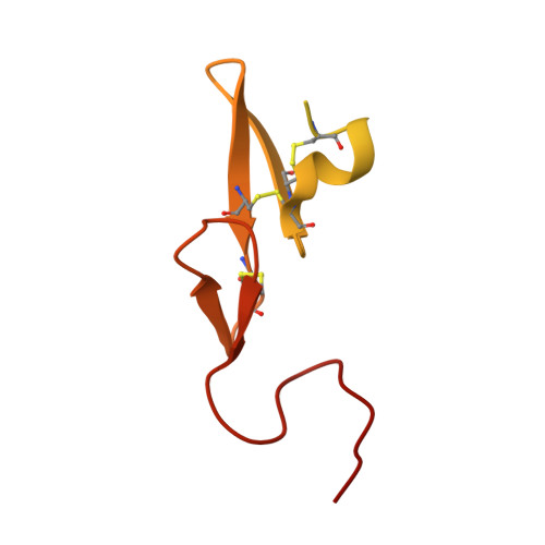
Crystal structures of human factor Xa complexed with potent inhibitors.

Maignan, S.Guilloteau, J.P.Pouzieux, S.Choi-Sledeski, Y.M.Becker, M.R.Klein, S.I.Ewing, W.R.Pauls, H.W.Spada, A.P.Mikol, V.

(2000) J Med Chem 43: 3226-3232

  • DOI: https://doi.org/10.1021/jm000940u
  • Primary Citation of Related Structures:  
    1EZQ, 1F0R, 1F0S, 1F0T, 1F0U

  • PubMed Abstract: 

    Involved in the coagulation cascade, factor Xa (FXa) is a serine protease which has received great interest as a potential target for the development of new antithrombotics. Although there is a great wealth of structural data on thrombin complexes, few structures of ligand/FXa complexes have been reported, presumably because of the difficulty in growing crystals. Reproducible crystallization conditions for human des-Gla1-45 coagulation FXa have been found. This has led to an improvement in the diffraction quality of the crystals (about 2.1 A) when compared to the previously reported forms (2.3-2.8 A) thus providing a suitable platform for a structure-based drug design approach. A series of crystal structures of noncovalent inhibitors complexed with FXa have been determined, three of which are presented herein. These include compounds containing the benzamidine moiety and surrogates of the basic group. The benzamidine-containing compound binds in a canonical fashion typical of synthetic serine protease inhibitors. On the contrary, molecules that contain surrogates of the benzamidine group do not make direct hydrogen-bonding interactions with the carboxylate of Asp189 at the bottom of the S1 pocket. The structural data provide a likely explanation for the specificity of these inhibitors and a great aid in the design of bioavailable potent FXa inhibitors.


  • Organizational Affiliation

    Department of Structural Biology, Aventis Pharma, 13, Quai J. Guesde, F-94403 Vitry/Seine, France.


Macromolecules
Find similar proteins by:  (by identity cutoff)  |  3D Structure
Entity ID: 1
MoleculeChains Sequence LengthOrganismDetailsImage
COAGULATION FACTOR XA254Homo sapiensMutation(s): 0 
EC: 3.4.21.6
UniProt & NIH Common Fund Data Resources
Find proteins for P00742 (Homo sapiens)
Explore P00742 
Go to UniProtKB:  P00742
PHAROS:  P00742
GTEx:  ENSG00000126218 
Entity Groups  
Sequence Clusters30% Identity50% Identity70% Identity90% Identity95% Identity100% Identity
UniProt GroupP00742
Sequence Annotations
Expand
  • Reference Sequence
Find similar proteins by:  (by identity cutoff)  |  3D Structure
Entity ID: 2
MoleculeChains Sequence LengthOrganismDetailsImage
COAGULATION FACTOR XA134Homo sapiensMutation(s): 0 
EC: 3.4.21.6
UniProt & NIH Common Fund Data Resources
Find proteins for P00742 (Homo sapiens)
Explore P00742 
Go to UniProtKB:  P00742
PHAROS:  P00742
GTEx:  ENSG00000126218 
Entity Groups  
Sequence Clusters30% Identity50% Identity70% Identity90% Identity95% Identity100% Identity
UniProt GroupP00742
Sequence Annotations
Expand
  • Reference Sequence
Small Molecules
Ligands 2 Unique
IDChains Name / Formula / InChI Key2D Diagram3D Interactions
815
Query on 815

Download Ideal Coordinates CCD File 
D [auth A]THIENO[3,2-B]PYRIDINE-2-SULFONIC ACID [1-(1-AMINO-ISOQUINOLIN-7-YLMETHYL)-2-OXO-PYRROLDIN-3-YL]-AMIDE
C21 H19 N5 O3 S2
NVKDOURNRJCKJE-INIZCTEOSA-N
CA
Query on CA

Download Ideal Coordinates CCD File 
C [auth A]CALCIUM ION
Ca
BHPQYMZQTOCNFJ-UHFFFAOYSA-N
Binding Affinity Annotations 
IDSourceBinding Affinity
815 Binding MOAD:  1F0R Ki: 22 (nM) from 1 assay(s)
BindingDB:  1F0R Ki: 22 (nM) from 1 assay(s)
PDBBind:  1F0R Ki: 22 (nM) from 1 assay(s)
Experimental Data & Validation

Experimental Data

  • Method: X-RAY DIFFRACTION
  • Resolution: 2.10 Å
  • R-Value Free: 0.263 
  • R-Value Work: 0.216 
  • R-Value Observed: 0.216 
  • Space Group: P 21 21 21
Unit Cell:
Length ( Å )Angle ( ˚ )
a = 56.51α = 90
b = 72.228β = 90
c = 77.912γ = 90
Software Package:
Software NamePurpose
MOSFLMdata reduction
SCALAdata scaling
X-PLORmodel building
X-PLORrefinement
CCP4data scaling
X-PLORphasing

Structure Validation

View Full Validation Report


Ligand Structure Quality Assessment 


Entry History 

Deposition Data

Revision History  (Full details and data files)

  • Version 1.0: 2000-09-20
    Type: Initial release
  • Version 1.1: 2008-04-27
    Changes: Version format compliance
  • Version 1.2: 2011-07-13
    Changes: Version format compliance
  • Version 1.3: 2024-03-13
    Changes: Data collection, Database references, Derived calculations, Source and taxonomy, Structure summary