7Z0D

Crystal structure of the L state of bacteriorhodopsin at 1.20 Angstrom resolution


Experimental Data Snapshot

  • Method: X-RAY DIFFRACTION
  • Resolution: 1.20 Å
  • R-Value Free: 0.174 
  • R-Value Work: 0.154 
  • R-Value Observed: 0.155 

wwPDB Validation   3D Report Full Report


Ligand Structure Quality Assessment 


This is version 1.3 of the entry. See complete history


Literature

True-atomic-resolution insights into the structure and functional role of linear chains and low-barrier hydrogen bonds in proteins.

Borshchevskiy, V.Kovalev, K.Round, E.Efremov, R.Astashkin, R.Bourenkov, G.Bratanov, D.Balandin, T.Chizhov, I.Baeken, C.Gushchin, I.Kuzmin, A.Alekseev, A.Rogachev, A.Willbold, D.Engelhard, M.Bamberg, E.Buldt, G.Gordeliy, V.

(2022) Nat Struct Mol Biol 29: 440-450

  • DOI: https://doi.org/10.1038/s41594-022-00762-2
  • Primary Citation of Related Structures:  
    7Z09, 7Z0A, 7Z0C, 7Z0D, 7Z0E

  • PubMed Abstract: 

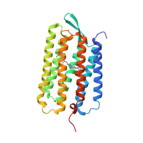
    Hydrogen bonds are fundamental to the structure and function of biological macromolecules and have been explored in detail. The chains of hydrogen bonds (CHBs) and low-barrier hydrogen bonds (LBHBs) were proposed to play essential roles in enzyme catalysis and proton transport. However, high-resolution structural data from CHBs and LBHBs is limited. The challenge is that their 'visualization' requires ultrahigh-resolution structures of the ground and functionally important intermediate states to identify proton translocation events and perform their structural assignment. Our true-atomic-resolution structures of the light-driven proton pump bacteriorhodopsin, a model in studies of proton transport, show that CHBs and LBHBs not only serve as proton pathways, but also are indispensable for long-range communications, signaling and proton storage in proteins. The complete picture of CHBs and LBHBs discloses their multifunctional roles in providing protein functions and presents a consistent picture of proton transport and storage resolving long-standing debates and controversies.


  • Organizational Affiliation

    Institute of Biological Information Processing (IBI-7: Structural Biochemistry), Forschungszentrum Jülich, Jülich, Germany.


Macromolecules
Find similar proteins by:  (by identity cutoff)  |  3D Structure
Entity ID: 1
MoleculeChains Sequence LengthOrganismDetailsImage
Bacteriorhodopsin248Halobacterium salinarumMutation(s): 0 
Membrane Entity: Yes 
UniProt
Find proteins for P02945 (Halobacterium salinarum (strain ATCC 700922 / JCM 11081 / NRC-1))
Explore P02945 
Go to UniProtKB:  P02945
Entity Groups  
Sequence Clusters30% Identity50% Identity70% Identity90% Identity95% Identity100% Identity
UniProt GroupP02945
Sequence Annotations
Expand
  • Reference Sequence
Small Molecules
Ligands 3 Unique
IDChains Name / Formula / InChI Key2D Diagram3D Interactions
OLC
Query on OLC

Download Ideal Coordinates CCD File 
D [auth A],
L [auth A],
X [auth A]
(2R)-2,3-dihydroxypropyl (9Z)-octadec-9-enoate
C21 H40 O4
RZRNAYUHWVFMIP-GDCKJWNLSA-N
LFA
Query on LFA

Download Ideal Coordinates CCD File 
AA [auth A]
B [auth A]
BA [auth A]
C [auth A]
E [auth A]
AA [auth A],
B [auth A],
BA [auth A],
C [auth A],
E [auth A],
F [auth A],
G [auth A],
H [auth A],
I [auth A],
J [auth A],
K [auth A],
M [auth A],
N [auth A],
O [auth A],
P [auth A],
Q [auth A],
R [auth A],
S [auth A],
T [auth A],
U [auth A],
V [auth A],
W [auth A],
Y [auth A]
EICOSANE
C20 H42
CBFCDTFDPHXCNY-UHFFFAOYSA-N
OLA
Query on OLA

Download Ideal Coordinates CCD File 
Z [auth A]OLEIC ACID
C18 H34 O2
ZQPPMHVWECSIRJ-KTKRTIGZSA-N
Modified Residues  1 Unique
IDChains TypeFormula2D DiagramParent
LYR
Query on LYR
A
L-PEPTIDE LINKINGC26 H42 N2 O2LYS
Experimental Data & Validation

Experimental Data

  • Method: X-RAY DIFFRACTION
  • Resolution: 1.20 Å
  • R-Value Free: 0.174 
  • R-Value Work: 0.154 
  • R-Value Observed: 0.155 
  • Space Group: P 63
Unit Cell:
Length ( Å )Angle ( ˚ )
a = 60.92α = 90
b = 60.92β = 90
c = 110.18γ = 120
Software Package:
Software NamePurpose
XSCALEdata scaling
PHENIXrefinement
PDB_EXTRACTdata extraction
XDSdata reduction
MOLREPphasing

Structure Validation

View Full Validation Report


Ligand Structure Quality Assessment 


Entry History & Funding Information

Deposition Data


Funding OrganizationLocationGrant Number
French Infrastructure for Integrated Structural Biology (FRISBI)FranceANR-10-INBS-05-02
Russian Foundation for Basic ResearchRussian Federation18-02-40020
Ministry of Science and Higher Education of the Russian FederationRussian Federation075-15-2021-1354
Russian Science FoundationRussian Federation21-64-00018
Ministry of Science and Higher Education of the Russian FederationRussian Federation075-00958-21-05/730000F.99.1.BV10AA00006
Ministry of Science and Higher Education of the Russian FederationRussian Federation075-00337-20-03/FSMG-2020-0003

Revision History  (Full details and data files)

  • Version 1.0: 2022-05-04
    Type: Initial release
  • Version 1.1: 2022-05-11
    Changes: Database references
  • Version 1.2: 2022-06-01
    Changes: Database references
  • Version 1.3: 2024-01-31
    Changes: Data collection, Refinement description