7VPP

Structures of a deltacoronavirus spike protein bound to porcine and human receptors indicate the risk of virus adaptation to humans


Experimental Data Snapshot

  • Method: X-RAY DIFFRACTION
  • Resolution: 2.69 Å
  • R-Value Free: 0.230 
  • R-Value Work: 0.198 
  • R-Value Observed: 0.200 

wwPDB Validation   3D Report Full Report


Ligand Structure Quality Assessment 


This is version 1.2 of the entry. See complete history


Literature

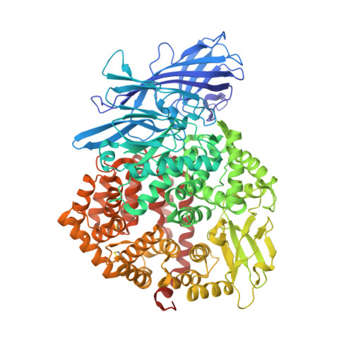
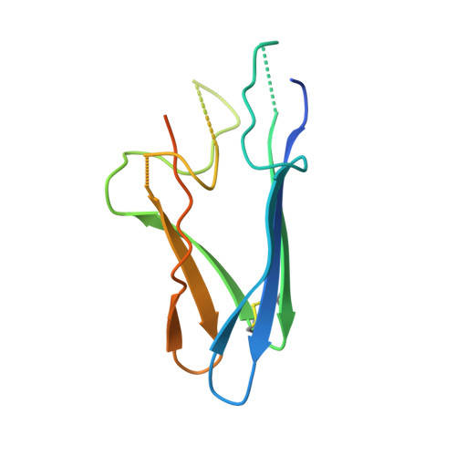
Structures of a deltacoronavirus spike protein bound to porcine and human receptors.

Ji, W.Peng, Q.Fang, X.Li, Z.Li, Y.Xu, C.Zhao, S.Li, J.Chen, R.Mo, G.Wei, Z.Xu, Y.Li, B.Zhang, S.

(2022) Nat Commun 13: 1467-1467

  • DOI: https://doi.org/10.1038/s41467-022-29062-5
  • Primary Citation of Related Structures:  
    7VPP, 7VPQ

  • PubMed Abstract: 

    Porcine deltacoronavirus (PDCoV) can experimentally infect a variety of animals. Human infection by PDCoV has also been reported. Consistently, PDCoV can use aminopeptidase N (APN) from different host species as receptors to enter cells. To understand this broad receptor usage and interspecies transmission of PDCoV, we determined the crystal structures of the receptor binding domain (RBD) of PDCoV spike protein bound to human APN (hAPN) and porcine APN (pAPN), respectively. The structures of the two complexes exhibit high similarity. PDCoV RBD binds to common regions on hAPN and pAPN, which are different from the sites engaged by two alphacoronaviruses: HCoV-229E and porcine respiratory coronavirus (PRCoV). Based on structure guided mutagenesis, we identified conserved residues on hAPN and pAPN that are essential for PDCoV binding and infection. We report the detailed mechanism for how a deltacoronavirus recognizes homologous receptors and provide insights into the cross-species transmission of PDCoV.


  • Organizational Affiliation

    College of Life Sciences, Nanjing Agricultural University, Nanjing, 210095, China.


Macromolecules
Find similar proteins by:  (by identity cutoff)  |  3D Structure
Entity ID: 1
MoleculeChains Sequence LengthOrganismDetailsImage
Aminopeptidase
A, C
911Sus scrofaMutation(s): 0 
Gene Names: ANPEPpAPN
EC: 3.4.11
UniProt
Find proteins for P15145 (Sus scrofa)
Explore P15145 
Go to UniProtKB:  P15145
Entity Groups  
Sequence Clusters30% Identity50% Identity70% Identity90% Identity95% Identity100% Identity
UniProt GroupP15145
Sequence Annotations
Expand
  • Reference Sequence
Find similar proteins by:  (by identity cutoff)  |  3D Structure
Entity ID: 2
MoleculeChains Sequence LengthOrganismDetailsImage
Spike protein127Porcine deltacoronavirusMutation(s): 0 
UniProt
Find proteins for A0A4P8D758 (Porcine deltacoronavirus)
Explore A0A4P8D758 
Go to UniProtKB:  A0A4P8D758
Entity Groups  
Sequence Clusters30% Identity50% Identity70% Identity90% Identity95% Identity100% Identity
UniProt GroupA0A4P8D758
Sequence Annotations
Expand
  • Reference Sequence
Oligosaccharides

Help

Entity ID: 3
MoleculeChains Length2D Diagram Glycosylation3D Interactions
2-acetamido-2-deoxy-beta-D-glucopyranose-(1-4)-2-acetamido-2-deoxy-beta-D-glucopyranose
D, E, F, G, H
D, E, F, G, H, I, J, K, M
2N-Glycosylation
Glycosylation Resources
GlyTouCan:  G42666HT
GlyCosmos:  G42666HT
GlyGen:  G42666HT
Entity ID: 4
MoleculeChains Length2D Diagram Glycosylation3D Interactions
2-acetamido-2-deoxy-beta-D-glucopyranose-(1-3)-2-acetamido-2-deoxy-beta-D-glucopyranose
L
2N-Glycosylation
Glycosylation Resources
GlyTouCan:  G79924RM
GlyCosmos:  G79924RM
GlyGen:  G79924RM
Small Molecules
Ligands 3 Unique
IDChains Name / Formula / InChI Key2D Diagram3D Interactions
NAG (Subject of Investigation/LOI)
Query on NAG

Download Ideal Coordinates CCD File 
R [auth A],
S [auth A],
T [auth B],
X [auth C],
Y [auth C]
2-acetamido-2-deoxy-beta-D-glucopyranose
C8 H15 N O6
OVRNDRQMDRJTHS-FMDGEEDCSA-N
GOL (Subject of Investigation/LOI)
Query on GOL

Download Ideal Coordinates CCD File 
O [auth A],
P [auth A],
Q [auth A],
V [auth C],
W [auth C]
GLYCEROL
C3 H8 O3
PEDCQBHIVMGVHV-UHFFFAOYSA-N
ZN (Subject of Investigation/LOI)
Query on ZN

Download Ideal Coordinates CCD File 
N [auth A],
U [auth C]
ZINC ION
Zn
PTFCDOFLOPIGGS-UHFFFAOYSA-N
Experimental Data & Validation

Experimental Data

  • Method: X-RAY DIFFRACTION
  • Resolution: 2.69 Å
  • R-Value Free: 0.230 
  • R-Value Work: 0.198 
  • R-Value Observed: 0.200 
  • Space Group: P 41 21 2
Unit Cell:
Length ( Å )Angle ( ˚ )
a = 186.643α = 90
b = 186.643β = 90
c = 173.823γ = 90
Software Package:
Software NamePurpose
PHENIXrefinement
PDB_EXTRACTdata extraction
XDSdata reduction
XDSdata scaling
PHASERphasing

Structure Validation

View Full Validation Report


Ligand Structure Quality Assessment 


Entry History & Funding Information

Deposition Data


Funding OrganizationLocationGrant Number
National Natural Science Foundation of China (NSFC)China32000659

Revision History  (Full details and data files)

  • Version 1.0: 2022-03-16
    Type: Initial release
  • Version 1.1: 2022-09-28
    Changes: Database references
  • Version 1.2: 2023-11-29
    Changes: Data collection, Refinement description