7VOC

The crystal structure of a Radical SAM Enzyme BlsE involved in the Biosynthesis of Blasticidin S


Experimental Data Snapshot

  • Method: X-RAY DIFFRACTION
  • Resolution: 2.62 Å
  • R-Value Free: 0.234 
  • R-Value Work: 0.214 
  • R-Value Observed: 0.215 

wwPDB Validation   3D Report Full Report


Ligand Structure Quality Assessment 


This is version 1.1 of the entry. See complete history


Literature

Radical S -Adenosyl Methionine Enzyme BlsE Catalyzes a Radical-Mediated 1,2-Diol Dehydration during the Biosynthesis of Blasticidin S.

Lee, Y.H.Hou, X.Chen, R.Feng, J.Liu, X.Ruszczycky, M.W.Gao, J.M.Wang, B.Zhou, J.Liu, H.W.

(2022) J Am Chem Soc 144: 4478-4486

  • DOI: https://doi.org/10.1021/jacs.1c12010
  • Primary Citation of Related Structures:  
    7VOB, 7VOC

  • PubMed Abstract: 

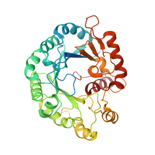
    The biosynthesis of blasticidin S has drawn attention due to the participation of the radical S -adenosyl methionine (SAM) enzyme BlsE. The original assignment of BlsE as a radical-mediated, redox-neutral decarboxylase is unusual because this reaction appears to serve no biosynthetic purpose and would need to be reversed by a subsequent carboxylation step. Furthermore, with the exception of BlsE, all other radical SAM decarboxylases reported to date are oxidative in nature. Careful analysis of the BlsE reaction, however, demonstrates that BlsE is not a decarboxylase but instead a lyase that catalyzes the dehydration of cytosylglucuronic acid (CGA) to form cytosyl-4'-keto-3'-deoxy-d-glucuronic acid, which can rapidly decarboxylate nonenzymatically in vitro . Analysis of substrate isotopologs, fluorinated analogues, as well as computational models based on X-ray crystal structures of the BlsE·SAM (2.09 Å) and BlsE·SAM·CGA (2.62 Å) complexes suggests that BlsE catalysis likely proceeds via direct elimination of water from the CGA C4' α-hydroxyalkyl radical as opposed to 1,2-migration of the C3'-hydroxyl prior to dehydration. Biosynthetic and mechanistic implications of the revised assignment of BlsE are discussed.


  • Organizational Affiliation

    Department of Chemistry, University of Texas at Austin, Austin, Texas 78712, United States.


Macromolecules
Find similar proteins by:  (by identity cutoff)  |  3D Structure
Entity ID: 1
MoleculeChains Sequence LengthOrganismDetailsImage
Cytosylglucuronate decarboxylaseA [auth C]364Streptomyces griseochromogenesMutation(s): 0 
Gene Names: AVL59_19980
UniProt
Find proteins for A0A1B1AYF2 (Streptomyces griseochromogenes)
Explore A0A1B1AYF2 
Go to UniProtKB:  A0A1B1AYF2
Entity Groups  
Sequence Clusters30% Identity50% Identity70% Identity90% Identity95% Identity100% Identity
UniProt GroupA0A1B1AYF2
Sequence Annotations
Expand
  • Reference Sequence
Small Molecules
Experimental Data & Validation

Experimental Data

  • Method: X-RAY DIFFRACTION
  • Resolution: 2.62 Å
  • R-Value Free: 0.234 
  • R-Value Work: 0.214 
  • R-Value Observed: 0.215 
  • Space Group: I 41 2 2
Unit Cell:
Length ( Å )Angle ( ˚ )
a = 163.92α = 90
b = 163.92β = 90
c = 158.81γ = 90
Software Package:
Software NamePurpose
PHENIXrefinement
PHENIXrefinement
xia2data reduction
xia2data scaling
PHASERphasing

Structure Validation

View Full Validation Report


Ligand Structure Quality Assessment 


Entry History & Funding Information

Deposition Data


Funding OrganizationLocationGrant Number
Not funded--

Revision History  (Full details and data files)

  • Version 1.0: 2022-05-04
    Type: Initial release
  • Version 1.1: 2023-11-29
    Changes: Data collection, Refinement description