


Crystal structure of the regulatory domain of AceR in Acinetobacter baumannii
Chi, P., Guo, L.To be published.
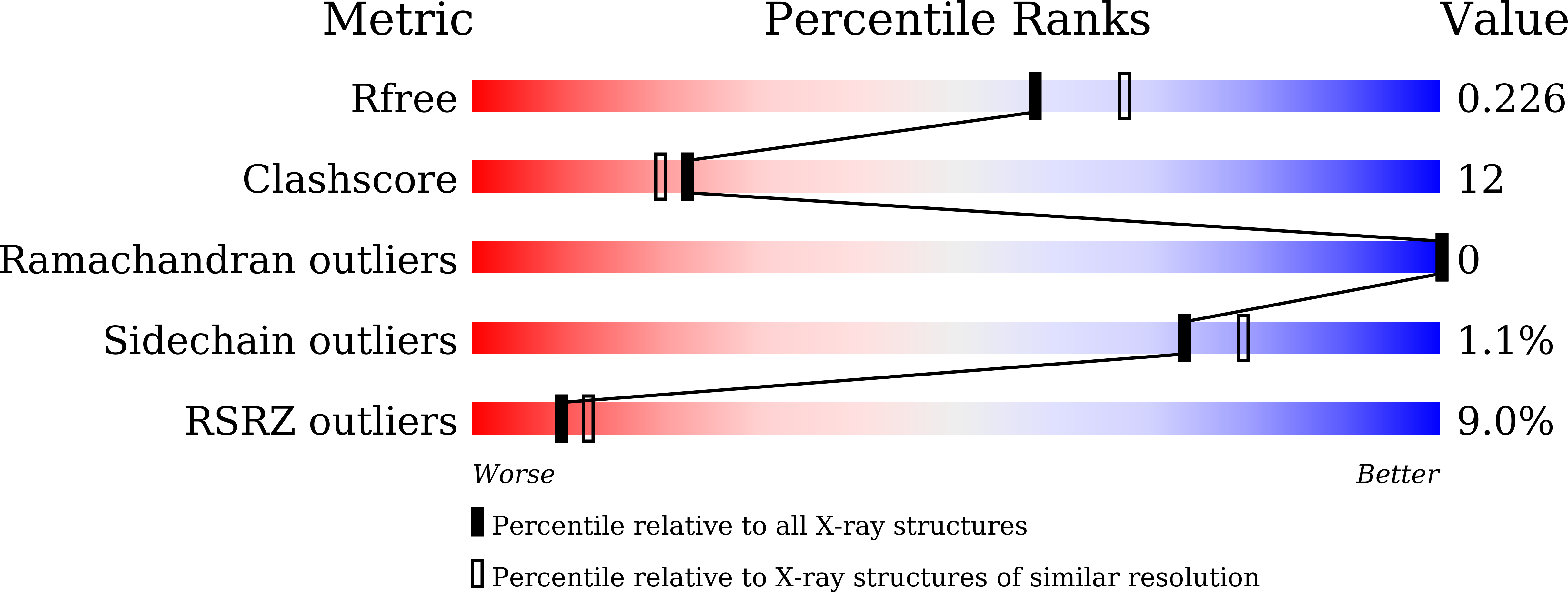
Experimental Data Snapshot
wwPDB Validation 3D Report Full Report
Entity ID: 1 | |||||
|---|---|---|---|---|---|
| Molecule | Chains | Sequence Length | Organism | Details | Image |
| LysR family transcriptional regulator | 213 | Acinetobacter baumannii | Mutation(s): 0 Gene Names: AceR | 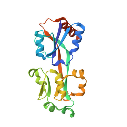 | |
UniProt | |||||
Find proteins for P0DUU5 (Acinetobacter baumannii (strain ATCC 17978 / CIP 53.77 / LMG 1025 / NCDC KC755 / 5377)) Explore P0DUU5 Go to UniProtKB: P0DUU5 | |||||
Entity Groups | |||||
| Sequence Clusters | 30% Identity50% Identity70% Identity90% Identity95% Identity100% Identity | ||||
| UniProt Group | P0DUU5 | ||||
Sequence AnnotationsExpand | |||||
| |||||
| Ligands 1 Unique | |||||
|---|---|---|---|---|---|
| ID | Chains | Name / Formula / InChI Key | 2D Diagram | 3D Interactions | |
| BR Query on BR | C [auth A], D [auth A], E [auth B], F [auth B], G [auth B] | BROMIDE ION Br CPELXLSAUQHCOX-UHFFFAOYSA-M |  | ||
| Length ( Å ) | Angle ( ˚ ) |
|---|---|
| a = 125.215 | α = 90 |
| b = 125.215 | β = 90 |
| c = 61.58 | γ = 120 |
| Software Name | Purpose |
|---|---|
| PHENIX | refinement |
| SCALEPACK | data scaling |
| PDB_EXTRACT | data extraction |
| HKL-3000 | data reduction |
| CRANK2 | phasing |
RCSB PDB (citation) is hosted by
RCSB PDB is a member of the

