7AV7

Crystal structure of S-nitrosylated nitrosoglutathione reductase(GSNOR)from Chlamydomonas reinhardtii, in complex with NAD+


Experimental Data Snapshot

  • Method: X-RAY DIFFRACTION
  • Resolution: 2.90 Å
  • R-Value Free: 0.257 
  • R-Value Work: 0.180 
  • R-Value Observed: 0.184 

wwPDB Validation   3D Report Full Report


Ligand Structure Quality Assessment 


This is version 1.1 of the entry. See complete history


Literature

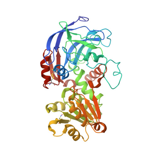
Structural and functional insights into nitrosoglutathione reductase from Chlamydomonas reinhardtii.

Tagliani, A.Rossi, J.Marchand, C.H.De Mia, M.Tedesco, D.Gurrieri, L.Meloni, M.Falini, G.Trost, P.Lemaire, S.D.Fermani, S.Zaffagnini, M.

(2020) Redox Biol 38: 101806-101806

  • DOI: https://doi.org/10.1016/j.redox.2020.101806
  • Primary Citation of Related Structures:  
    7AAS, 7AAU, 7AV7

  • PubMed Abstract: 

    Protein S-nitrosylation plays a fundamental role in cell signaling and nitrosoglutathione (GSNO) is considered as the main nitrosylating signaling molecule. Enzymatic systems controlling GSNO homeostasis are thus crucial to indirectly control the formation of protein S-nitrosothiols. GSNO reductase (GSNOR) is the key enzyme controlling GSNO levels by catalyzing its degradation in the presence of NADH. Here, we found that protein extracts from the microalga Chlamydomonas reinhardtii catabolize GSNO via two enzymatic systems having specific reliance on NADPH or NADH and different biochemical features. Scoring the Chlamydomonas genome for orthologs of known plant GSNORs, we found two genes encoding for putative and almost identical GSNOR isoenzymes. One of the two, here named CrGSNOR1, was heterologously expressed and purified. Its kinetic properties were determined and the three-dimensional structures of the apo-, NAD + - and NAD + /GSNO-forms were solved. These analyses revealed that CrGSNOR1 has a strict specificity towards GSNO and NADH, and a conserved folding with respect to other plant GSNORs. The catalytic zinc ion, however, showed an unexpected variability of the coordination environment. Furthermore, we evaluated the catalytic response of CrGSNOR1 to thermal denaturation, thiol-modifying agents and oxidative modifications as well as the reactivity and position of accessible cysteines. Despite being a cysteine-rich protein, CrGSNOR1 contains only two solvent-exposed/reactive cysteines. Oxidizing and nitrosylating treatments have null or limited effects on CrGSNOR1 activity and folding, highlighting a certain resistance of the algal enzyme to redox modifications. The molecular mechanisms and structural features underlying the response to thiol-based modifications are discussed.


  • Organizational Affiliation

    Department of Pharmacy and Biotechnologies, University of Bologna, I-40126, Bologna, Italy; CNRS, Sorbonne Université, Institut de Biologie Physico-Chimique, UMR8226, F-75005, Paris, France.


Macromolecules
Find similar proteins by:  (by identity cutoff)  |  3D Structure
Entity ID: 1
MoleculeChains Sequence LengthOrganismDetailsImage
S-(hydroxymethyl)glutathione dehydrogenase
A, B, C, D, E
A, B, C, D, E, F
378Chlamydomonas reinhardtiiMutation(s): 0 
Gene Names: CHLRE_12g543400v5
EC: 1.1.1.284
UniProt
Find proteins for A0A2K3D6R4 (Chlamydomonas reinhardtii)
Explore A0A2K3D6R4 
Go to UniProtKB:  A0A2K3D6R4
Entity Groups  
Sequence Clusters30% Identity50% Identity70% Identity90% Identity95% Identity100% Identity
UniProt GroupA0A2K3D6R4
Sequence Annotations
Expand
  • Reference Sequence
Small Molecules
Ligands 3 Unique
IDChains Name / Formula / InChI Key2D Diagram3D Interactions
NAD (Subject of Investigation/LOI)
Query on NAD

Download Ideal Coordinates CCD File 
I [auth A]
L [auth B]
O [auth C]
R [auth D]
V [auth E]
I [auth A],
L [auth B],
O [auth C],
R [auth D],
V [auth E],
Y [auth F]
NICOTINAMIDE-ADENINE-DINUCLEOTIDE
C21 H27 N7 O14 P2
BAWFJGJZGIEFAR-NNYOXOHSSA-N
ZN (Subject of Investigation/LOI)
Query on ZN

Download Ideal Coordinates CCD File 
G [auth A]
H [auth A]
J [auth B]
K [auth B]
M [auth C]
G [auth A],
H [auth A],
J [auth B],
K [auth B],
M [auth C],
N [auth C],
P [auth D],
Q [auth D],
T [auth E],
U [auth E],
W [auth F],
X [auth F]
ZINC ION
Zn
PTFCDOFLOPIGGS-UHFFFAOYSA-N
CL
Query on CL

Download Ideal Coordinates CCD File 
S [auth D]CHLORIDE ION
Cl
VEXZGXHMUGYJMC-UHFFFAOYSA-M
Modified Residues  1 Unique
IDChains TypeFormula2D DiagramParent
SNC
Query on SNC
A, B, C, D, E
A, B, C, D, E, F
L-PEPTIDE LINKINGC3 H6 N2 O3 SCYS
Experimental Data & Validation

Experimental Data

  • Method: X-RAY DIFFRACTION
  • Resolution: 2.90 Å
  • R-Value Free: 0.257 
  • R-Value Work: 0.180 
  • R-Value Observed: 0.184 
  • Space Group: P 21 21 21
Unit Cell:
Length ( Å )Angle ( ˚ )
a = 78.386α = 90
b = 143.294β = 90
c = 206.271γ = 90
Software Package:
Software NamePurpose
SCALAdata scaling
PHENIXrefinement
PDB_EXTRACTdata extraction
XDSdata reduction
PHASERphasing

Structure Validation

View Full Validation Report


Ligand Structure Quality Assessment 


Entry History & Funding Information

Deposition Data


Funding OrganizationLocationGrant Number
Other governmentItalyAlma Idea 2017
Agence Nationale de la Recherche (ANR)France17-CE05-0001

Revision History  (Full details and data files)

  • Version 1.0: 2020-12-30
    Type: Initial release
  • Version 1.1: 2024-01-31
    Changes: Data collection, Database references, Refinement description