6Z0Z

Human wtSTING in complex with 3',3'-c-(2'FdAMP-2'FdAMP)


Experimental Data Snapshot

  • Method: X-RAY DIFFRACTION
  • Resolution: 2.50 Å
  • R-Value Free: 0.227 
  • R-Value Work: 0.191 
  • R-Value Observed: 0.193 

wwPDB Validation   3D Report Full Report


Ligand Structure Quality Assessment 


This is version 1.1 of the entry. See complete history


Literature

Ligand Strain and Its Conformational Complexity Is a Major Factor in the Binding of Cyclic Dinucleotides to STING Protein.

Smola, M.Gutten, O.Dejmek, M.Kozisek, M.Evangelidis, T.Tehrani, Z.A.Novotna, B.Nencka, R.Birkus, G.Rulisek, L.Boura, E.

(2021) Angew Chem Int Ed Engl 60: 10172-10178

  • DOI: https://doi.org/10.1002/anie.202016805
  • Primary Citation of Related Structures:  
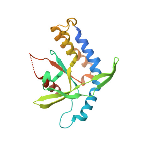
    6Y99, 6YDB, 6YDZ, 6YEA, 6Z0Z, 6Z15

  • PubMed Abstract: 

    STING (stimulator of interferon genes) is a key regulator of innate immunity that has recently been recognized as a promising drug target. STING is activated by cyclic dinucleotides (CDNs) which eventually leads to expression of type I interferons and other cytokines. Factors underlying the affinity of various CDN analogues are poorly understood. Herein, we correlate structural biology, isothermal calorimetry (ITC) and computational modeling to elucidate factors contributing to binding of six CDNs-three pairs of natural (ribo) and fluorinated (2'-fluororibo) 3',3'-CDNs. X-ray structural analyses of six {STING:CDN} complexes did not offer any explanation for the different affinities of the studied ligands. ITC showed entropy/enthalpy compensation up to 25 kcal mol -1 for this set of similar ligands. The higher affinities of fluorinated analogues are explained with help of computational methods by smaller loss of entropy upon binding and by smaller strain (free) energy.


  • Organizational Affiliation

    Gilead Sciences Research Centre at IOCB, Institute of Organic Chemistry and Biochemistry of the Czech Academy of Sciences, Flemingovo náměstí 2, 16610, Prague, Czech Republic.


Macromolecules
Find similar proteins by:  (by identity cutoff)  |  3D Structure
Entity ID: 1
MoleculeChains Sequence LengthOrganismDetailsImage
Stimulator of interferon protein204Homo sapiensMutation(s): 0 
Gene Names: STINGLOC340061hCG_1782396
UniProt & NIH Common Fund Data Resources
Find proteins for Q86WV6 (Homo sapiens)
Explore Q86WV6 
Go to UniProtKB:  Q86WV6
PHAROS:  Q86WV6
GTEx:  ENSG00000184584 
Entity Groups  
Sequence Clusters30% Identity50% Identity70% Identity90% Identity95% Identity100% Identity
UniProt GroupQ86WV6
Sequence Annotations
Expand
  • Reference Sequence
Small Molecules
Ligands 1 Unique
IDChains Name / Formula / InChI Key2D Diagram3D Interactions
M8T (Subject of Investigation/LOI)
Query on M8T

Download Ideal Coordinates CCD File 
B [auth A]2'-fluoro-,3',3'-c-di-AMP
C20 H22 F2 N10 O10 P2
LMPBRGUYJGJYOI-QPMBAPTHSA-N
Experimental Data & Validation

Experimental Data

  • Method: X-RAY DIFFRACTION
  • Resolution: 2.50 Å
  • R-Value Free: 0.227 
  • R-Value Work: 0.191 
  • R-Value Observed: 0.193 
  • Space Group: P 43 21 2
Unit Cell:
Length ( Å )Angle ( ˚ )
a = 67.174α = 90
b = 67.174β = 90
c = 136.047γ = 90
Software Package:
Software NamePurpose
XDSdata reduction
Aimlessdata scaling
PHASERphasing
PHENIXrefinement
PDB_EXTRACTdata extraction

Structure Validation

View Full Validation Report


Ligand Structure Quality Assessment 


Entry History 

Deposition Data

Revision History  (Full details and data files)

  • Version 1.0: 2021-05-19
    Type: Initial release
  • Version 1.1: 2024-01-24
    Changes: Data collection, Database references, Refinement description