6W1J

Cryo-EM structure of 5HT3A receptor in presence of Alosetron


Experimental Data Snapshot

  • Method: ELECTRON MICROSCOPY
  • Resolution: 2.92 Å
  • Aggregation State: PARTICLE 
  • Reconstruction Method: SINGLE PARTICLE 

wwPDB Validation   3D Report Full Report


This is version 1.0 of the entry. See complete history


Literature

High-resolution structures of multiple 5-HT 3A R-setron complexes reveal a novel mechanism of competitive inhibition.

Basak, S.Kumar, A.Ramsey, S.Gibbs, E.Kapoor, A.Filizola, M.Chakrapani, S.

(2020) Elife 9

  • DOI: https://doi.org/10.7554/eLife.57870
  • Primary Citation of Related Structures:  
    6W1J, 6W1M, 6W1Y

  • PubMed Abstract: 

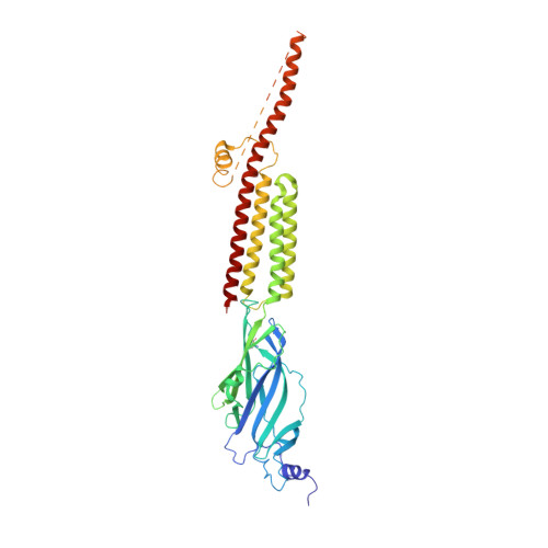
    Serotonin receptors (5-HT 3A R) play a crucial role in regulating gut movement, and are the principal target of setrons, a class of high-affinity competitive antagonists, used in the management of nausea and vomiting associated with radiation and chemotherapies. Structural insights into setron-binding poses and their inhibitory mechanisms are just beginning to emerge. Here, we present high-resolution cryo-EM structures of full-length 5-HT 3A R in complex with palonosetron, ondansetron, and alosetron. Molecular dynamic simulations of these structures embedded in a fully-hydrated lipid environment assessed the stability of ligand-binding poses and drug-target interactions over time. Together with simulation results of apo- and serotonin-bound 5-HT 3A R, the study reveals a distinct interaction fingerprint between the various setrons and binding-pocket residues that may underlie their diverse affinities. In addition, varying degrees of conformational change in the setron-5-HT 3A R structures, throughout the channel and particularly along the channel activation pathway, suggests a novel mechanism of competitive inhibition.


  • Organizational Affiliation

    Department of Physiology and Biophysics, Case Western Reserve University, Cleveland, United States.


Macromolecules
Find similar proteins by:  (by identity cutoff)  |  3D Structure
Entity ID: 1
MoleculeChains Sequence LengthOrganismDetailsImage
5-hydroxytryptamine receptor 3A
A, B, C, D, E
450Mus musculusMutation(s): 0 
Gene Names: Htr3a
Membrane Entity: Yes 
UniProt & NIH Common Fund Data Resources
Find proteins for P23979 (Mus musculus)
Explore P23979 
Go to UniProtKB:  P23979
IMPC:  MGI:96282
Entity Groups  
Sequence Clusters30% Identity50% Identity70% Identity90% Identity95% Identity100% Identity
UniProt GroupP23979
Sequence Annotations
Expand
  • Reference Sequence
Oligosaccharides

Help

Entity ID: 2
MoleculeChains Length2D Diagram Glycosylation3D Interactions
2-acetamido-2-deoxy-beta-D-glucopyranose-(4-4)-2-acetamido-2-deoxy-beta-D-glucopyranose
F, I, L, O, R
2N/AN-Glycosylation
Entity ID: 3
MoleculeChains Length2D Diagram Glycosylation3D Interactions
beta-D-mannopyranose-(1-4)-2-acetamido-2-deoxy-beta-D-glucopyranose-(1-4)-2-acetamido-2-deoxy-beta-D-glucopyranose
G, J, M, P, S
3N/AN-Glycosylation
Entity ID: 4
MoleculeChains Length2D Diagram Glycosylation3D Interactions
2-acetamido-2-deoxy-beta-D-glucopyranose-(1-4)-2-acetamido-2-deoxy-beta-D-glucopyranose
H, K, N, Q, T
2N/AN-Glycosylation
Experimental Data & Validation

Experimental Data

  • Method: ELECTRON MICROSCOPY
  • Resolution: 2.92 Å
  • Aggregation State: PARTICLE 
  • Reconstruction Method: SINGLE PARTICLE 
EM Software:
TaskSoftware PackageVersion
RECONSTRUCTIONRELION3.1
MODEL REFINEMENTPHENIX1.13-2998

Structure Validation

View Full Validation Report


Entry History & Funding Information

Deposition Data


Funding OrganizationLocationGrant Number
National Institutes of Health/National Institute of General Medical Sciences (NIH/NIGMS)United States1R01GM108921
National Institutes of Health/National Institute of General Medical Sciences (NIH/NIGMS)United States3R01GM108921-03S1

Revision History  (Full details and data files)

  • Version 1.0: 2021-01-13
    Type: Initial release