6VR6

Structure of ALDH9A1 complexed with NAD+ in space group P1


Experimental Data Snapshot

  • Method: X-RAY DIFFRACTION
  • Resolution: 2.50 Å
  • R-Value Free: 0.269 
  • R-Value Work: 0.213 
  • R-Value Observed: 0.214 

wwPDB Validation   3D Report Full Report

Currently 6VR6 does not have a validation slider image.


This is version 1.2 of the entry. See complete history


Literature

Inhibition, crystal structures, and in-solution oligomeric structure of aldehyde dehydrogenase 9A1.

Wyatt, J.W.Korasick, D.A.Qureshi, I.A.Campbell, A.C.Gates, K.S.Tanner, J.J.

(2020) Arch Biochem Biophys 691: 108477-108477

  • DOI: https://doi.org/10.1016/j.abb.2020.108477
  • Primary Citation of Related Structures:  
    6VR6, 6VWF

  • PubMed Abstract: 

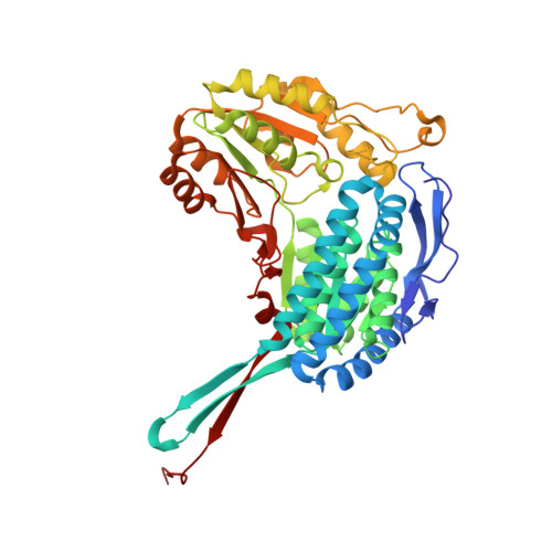
    Aldehyde dehydrogenase 9A1 (ALDH9A1) is a human enzyme that catalyzes the NAD + -dependent oxidation of the carnitine precursor 4-trimethylaminobutyraldehyde to 4-N-trimethylaminobutyrate. Here we show that the broad-spectrum ALDH inhibitor diethylaminobenzaldehyde (DEAB) reversibly inhibits ALDH9A1 in a time-dependent manner. Possible mechanisms of inhibition include covalent reversible inactivation involving the thiohemiacetal intermediate and slow, tight-binding inhibition. Two crystal structures of ALDH9A1 are reported, including the first of the enzyme complexed with NAD + . One of the structures reveals the active conformation of the enzyme, in which the Rossmann dinucleotide-binding domain is fully ordered and the inter-domain linker adopts the canonical β-hairpin observed in other ALDH structures. The oligomeric structure of ALDH9A1 was investigated using analytical ultracentrifugation, small-angle X-ray scattering, and negative stain electron microscopy. These data show that ALDH9A1 forms the classic ALDH superfamily dimer-of-dimers tetramer in solution. Our results suggest that the presence of an aldehyde substrate and NAD + promotes isomerization of the enzyme into the active conformation.


  • Organizational Affiliation

    Department of Chemistry, University of Missouri, Columbia, MO, 65211, United States.


Macromolecules
Find similar proteins by:  (by identity cutoff)  |  3D Structure
Entity ID: 1
MoleculeChains Sequence LengthOrganismDetailsImage
4-trimethylaminobutyraldehyde dehydrogenase
A, B, C, D, E
A, B, C, D, E, F, G, H
493Homo sapiensMutation(s): 0 
Gene Names: ALDH9A1ALDH4ALDH7ALDH9
EC: 1.2.1.47 (PDB Primary Data), 1.2.1.3 (PDB Primary Data), 1.2.1.19 (PDB Primary Data)
UniProt & NIH Common Fund Data Resources
Find proteins for P49189 (Homo sapiens)
Explore P49189 
Go to UniProtKB:  P49189
PHAROS:  P49189
GTEx:  ENSG00000143149 
Entity Groups  
Sequence Clusters30% Identity50% Identity70% Identity90% Identity95% Identity100% Identity
UniProt GroupP49189
Sequence Annotations
Expand
  • Reference Sequence
Experimental Data & Validation

Experimental Data

  • Method: X-RAY DIFFRACTION
  • Resolution: 2.50 Å
  • R-Value Free: 0.269 
  • R-Value Work: 0.213 
  • R-Value Observed: 0.214 
  • Space Group: P 1
Unit Cell:
Length ( Å )Angle ( ˚ )
a = 82.785α = 89.37
b = 90.297β = 84.04
c = 145.236γ = 73.87
Software Package:
Software NamePurpose
Aimlessdata scaling
PHENIXrefinement
PDB_EXTRACTdata extraction
XDSdata reduction
MOLREPphasing

Structure Validation

View Full Validation Report

Currently 6VR6 does not have a validation slider image.


Entry History & Funding Information

Deposition Data


Funding OrganizationLocationGrant Number
National Institutes of Health/National Institute of General Medical Sciences (NIH/NIGMS)United StatesR01GM093123
National Institutes of Health/National Institute of General Medical Sciences (NIH/NIGMS)United StatesR01GM065546

Revision History  (Full details and data files)

  • Version 1.0: 2020-08-05
    Type: Initial release
  • Version 1.1: 2020-08-26
    Changes: Database references
  • Version 1.2: 2023-10-11
    Changes: Data collection, Database references, Refinement description