


High resolution X-ray structure of glucagon and selected stereo-inversed analogs in novel crystallographic packing arrangement.
Mroz, P.A., Gonzalez-Gutierrez, G., DiMarchi, R.D.To be published.
Experimental Data Snapshot
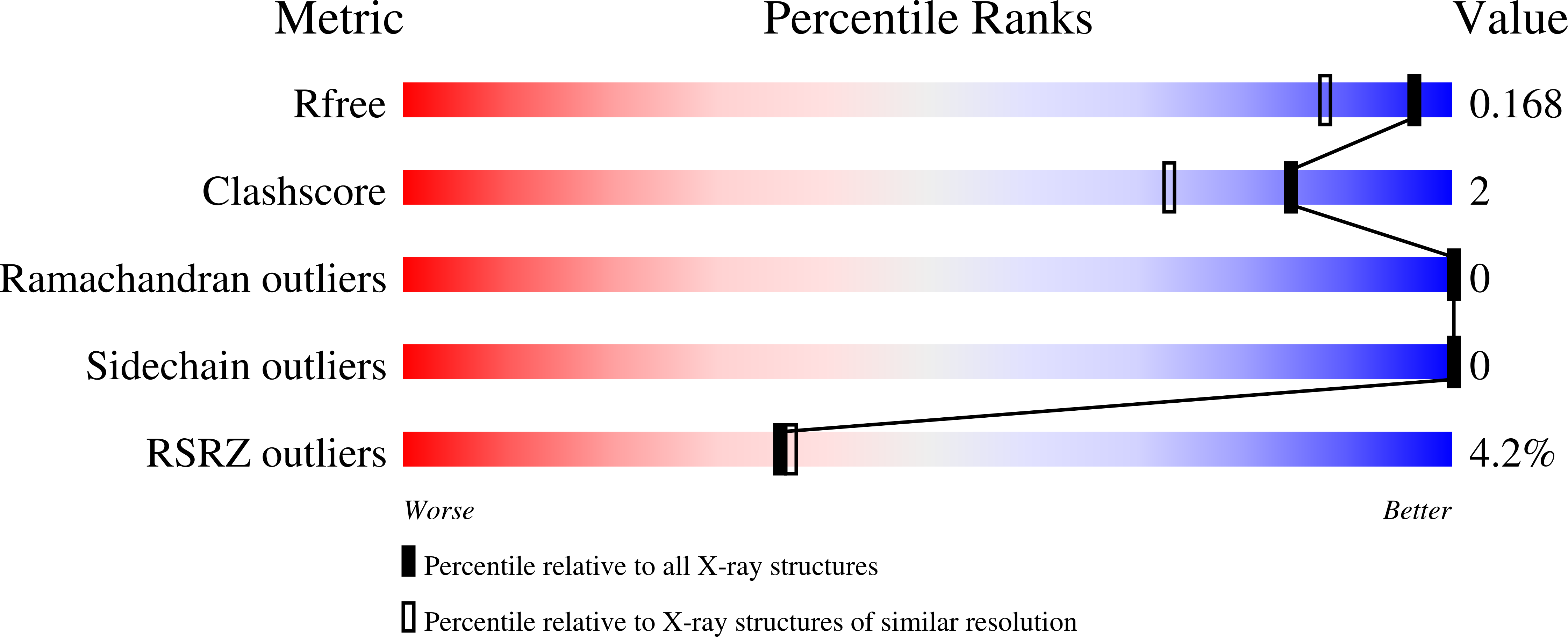
wwPDB Validation 3D Report Full Report
Entity ID: 1 | |||||
|---|---|---|---|---|---|
| Molecule | Chains | Sequence Length | Organism | Details | Image |
| Glucagon | 29 | Homo sapiens | Mutation(s): 1 |  | |
UniProt & NIH Common Fund Data Resources | |||||
Find proteins for P01275 (Homo sapiens) Explore P01275 Go to UniProtKB: P01275 | |||||
PHAROS: P01275 GTEx: ENSG00000115263 | |||||
Entity Groups | |||||
| Sequence Clusters | 30% Identity50% Identity70% Identity90% Identity95% Identity100% Identity | ||||
| UniProt Group | P01275 | ||||
Sequence AnnotationsExpand | |||||
| |||||
| Length ( Å ) | Angle ( ˚ ) |
|---|---|
| a = 40.168 | α = 90 |
| b = 40.168 | β = 90 |
| c = 37.794 | γ = 90 |
| Software Name | Purpose |
|---|---|
| PHENIX | refinement |
| XDS | data reduction |
| Aimless | data scaling |
| PHASER | phasing |
| Funding Organization | Location | Grant Number |
|---|---|---|
| Other government | United States | Indiana University Internal grant |
RCSB PDB (citation) is hosted by
RCSB PDB is a member of the

