6N2T

Ternary complex crystal structure of DNA polymerase Beta with 5-hydroxymethyl-dC (5-hmC) at the templating position


Experimental Data Snapshot

  • Method: X-RAY DIFFRACTION
  • Resolution: 2.60 Å
  • R-Value Free: 0.290 
  • R-Value Work: 0.198 
  • R-Value Observed: 0.208 

wwPDB Validation   3D Report Full Report


Ligand Structure Quality Assessment 


This is version 1.4 of the entry. See complete history


Literature

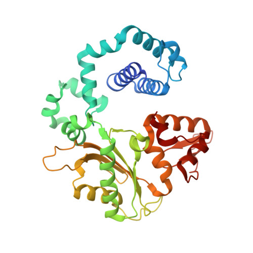
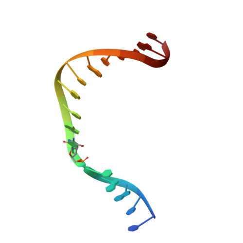
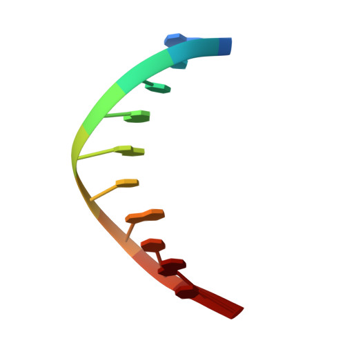
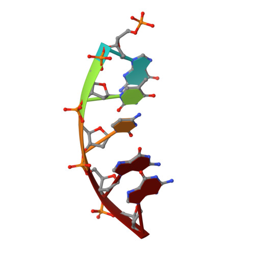
Molecular basis for the faithful replication of 5-methylcytosine and its oxidized forms by DNA polymerase beta.

Howard, M.J.Foley, K.G.Shock, D.D.Batra, V.K.Wilson, S.H.

(2019) J Biol Chem 294: 7194-7201

  • DOI: https://doi.org/10.1074/jbc.RA118.006809
  • Primary Citation of Related Structures:  
    6N2R, 6N2S, 6N2T

  • PubMed Abstract: 

    DNA methylation is an epigenetic mark that regulates gene expression in mammals. One method of methylation removal is through ten-eleven translocation-catalyzed oxidation and the base excision repair pathway. The iterative oxidation of 5-methylcytosine catalyzed by ten-eleven translocation enzymes produces three oxidized forms of cytosine: 5-hydroxmethylcytosine, 5-formylcytosine, and 5-carboxycytosine. The effect these modifications have on the efficiency and fidelity of the base excision repair pathway during the repair of opposing base damage, and in particular DNA polymerization, remains to be elucidated. Using kinetic assays, we show that the catalytic efficiency for the incorporation of dGTP catalyzed by human DNA polymerase β is not affected when 5-methylcytosine, 5-hydroxmethylcytosine, and 5-formylcytosine are in the DNA template. In contrast, the catalytic efficiency of dGTP insertion decreases ∼20-fold when 5-carboxycytosine is in the templating position, as compared with unmodified cytosine. However, DNA polymerase fidelity is unaltered when these modifications are in the templating position. Structural analysis reveals that the methyl, hydroxymethyl, and formyl modifications are easily accommodated within the polymerase active site. However, to accommodate the carboxyl modification, the phosphate backbone on the templating nucleotide shifts ∼2.5 Å to avoid a potential steric/repulsive clash. This altered conformation is stabilized by lysine 280, which makes a direct interaction with the carboxyl modification and the phosphate backbone of the templating strand. This work provides the molecular basis for the accommodation of epigenetic base modifications in a polymerase active site and suggests that these modifications are not mutagenically copied during base excision repair.


  • Organizational Affiliation

    From the Genome Integrity and Structural Biology Laboratory, NIEHS, National Institutes of Health, Research Triangle Park, North Carolina 27709.


Macromolecules

Find similar proteins by:  (by identity cutoff)  |  3D Structure
Entity ID: 1
MoleculeChains Sequence LengthOrganismDetailsImage
DNA polymerase beta335Homo sapiensMutation(s): 0 
Gene Names: POLB
EC: 2.7.7.7 (PDB Primary Data), 4.2.99 (PDB Primary Data)
UniProt & NIH Common Fund Data Resources
Find proteins for P06746 (Homo sapiens)
Explore P06746 
Go to UniProtKB:  P06746
PHAROS:  P06746
GTEx:  ENSG00000070501 
Entity Groups  
Sequence Clusters30% Identity50% Identity70% Identity90% Identity95% Identity100% Identity
UniProt GroupP06746
Sequence Annotations
Expand
  • Reference Sequence

Find similar nucleic acids by:  Sequence   |   3D Structure  

Entity ID: 2
MoleculeChains LengthOrganismImage
DNA (5'-D(*CP*CP*GP*AP*CP*(5HC)P*GP*CP*GP*CP*AP*TP*CP*AP*GP*C)-3')B [auth T]16Homo sapiens
Sequence Annotations
Expand
  • Reference Sequence

Find similar nucleic acids by:  Sequence   |   3D Structure  

Entity ID: 3
MoleculeChains LengthOrganismImage
DNA (5'-D(*GP*CP*TP*GP*AP*TP*GP*CP*GP*C)-3')C [auth P]10Homo sapiens
Sequence Annotations
Expand
  • Reference Sequence

Find similar nucleic acids by:  Sequence   |   3D Structure  

Entity ID: 4
MoleculeChains LengthOrganismImage
DNA (5'-D(P*GP*TP*CP*GP*G)-3')5Homo sapiens
Sequence Annotations
Expand
  • Reference Sequence
Experimental Data & Validation

Experimental Data

  • Method: X-RAY DIFFRACTION
  • Resolution: 2.60 Å
  • R-Value Free: 0.290 
  • R-Value Work: 0.198 
  • R-Value Observed: 0.208 
  • Space Group: P 1 21 1
Unit Cell:
Length ( Å )Angle ( ˚ )
a = 50.476α = 90
b = 79.089β = 106.61
c = 55.556γ = 90
Software Package:
Software NamePurpose
HKL-2000data reduction
HKL-2000data scaling
PHENIXrefinement
PDB_EXTRACTdata extraction

Structure Validation

View Full Validation Report


Ligand Structure Quality Assessment 


Entry History 

Deposition Data

Revision History  (Full details and data files)

  • Version 1.0: 2019-03-27
    Type: Initial release
  • Version 1.1: 2019-04-03
    Changes: Data collection, Database references
  • Version 1.2: 2019-05-01
    Changes: Data collection, Database references
  • Version 1.3: 2019-05-15
    Changes: Data collection, Database references
  • Version 1.4: 2023-10-11
    Changes: Data collection, Database references, Derived calculations, Refinement description