6GCD

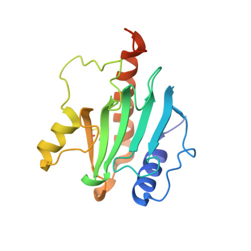
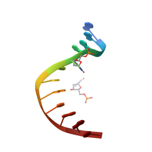
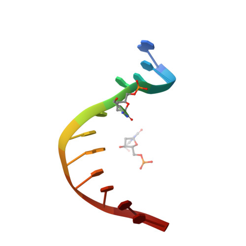
DNA binding domain of restriction endonuclease McrBC in complex with 5-hydroxymethylcytosine DNA


Experimental Data Snapshot

  • Method: X-RAY DIFFRACTION
  • Resolution: 1.80 Å
  • R-Value Free: 0.214 
  • R-Value Work: 0.166 
  • R-Value Observed: 0.171 

wwPDB Validation   3D Report Full Report


This is version 1.2 of the entry. See complete history


Literature

Recognition of modified cytosine variants by the DNA-binding domain of methyl-directed endonuclease McrBC.

Zagorskaite, E.Manakova, E.Sasnauskas, G.

(2018) FEBS Lett 592: 3335-3345

  • DOI: https://doi.org/10.1002/1873-3468.13244
  • Primary Citation of Related Structures:  
    6GCD, 6GCE, 6GCF

  • PubMed Abstract: 

    Cytosine modifications expand the information content of genomic DNA in both eukaryotes and prokaryotes, providing means for epigenetic regulation and self versus nonself discrimination. For example, the methyl-directed restriction endonuclease, McrBC, recognizes and cuts invading bacteriophage DNA containing 5-methylcytosine (5mC), 5-hydroxymethylcytosine (5hmC), and N4-methylcytosine (4mC), leaving the unmodified host DNA intact. Here, we present cocrystal structures of McrB-N bound to DNA oligoduplexes containing 5hmC, 5-formylcytosine (5fC), and 4mC, and characterize the relative affinity of McrB-N to various cytosine variants. We find that McrB-N flips out modified bases into a protein pocket and binds cytosine derivatives in the order of descending affinity: 4mC > 5mC > 5hmC ≫ 5fC. We also show that pocket mutations alter the relative preference of McrB-N to 5mC, 5hmC, and 4mC.


  • Organizational Affiliation

    Institute of Biotechnology, Vilnius University, Vilnius, Lithuania.


Macromolecules

Find similar proteins by:  (by identity cutoff)  |  3D Structure
Entity ID: 1
MoleculeChains Sequence LengthOrganismDetailsImage
5-methylcytosine-specific restriction enzyme B
A, B
170Escherichia coli K-12Mutation(s): 0 
Gene Names: mcrBrglBb4346JW5871
EC: 3.1.21
UniProt
Find proteins for P15005 (Escherichia coli (strain K12))
Explore P15005 
Go to UniProtKB:  P15005
Entity Groups  
Sequence Clusters30% Identity50% Identity70% Identity90% Identity95% Identity100% Identity
UniProt GroupP15005
Sequence Annotations
Expand
  • Reference Sequence

Find similar nucleic acids by:  Sequence   |   3D Structure  

Entity ID: 2
MoleculeChains LengthOrganismImage
DNA (5'-D(*GP*AP*GP*AP*(5HC)P*CP*GP*GP*TP*AP*GP*C)-3')12synthetic construct
Sequence Annotations
Expand
  • Reference Sequence

Find similar nucleic acids by:  Sequence   |   3D Structure  

Entity ID: 3
MoleculeChains LengthOrganismImage
DNA (5'-D(*GP*CP*TP*AP*(5HC)P*CP*GP*GP*TP*CP*TP*C)-3')12synthetic construct
Sequence Annotations
Expand
  • Reference Sequence
Small Molecules
Modified Residues  1 Unique
IDChains TypeFormula2D DiagramParent
CME
Query on CME
A, B
L-PEPTIDE LINKINGC5 H11 N O3 S2CYS
Experimental Data & Validation

Experimental Data

  • Method: X-RAY DIFFRACTION
  • Resolution: 1.80 Å
  • R-Value Free: 0.214 
  • R-Value Work: 0.166 
  • R-Value Observed: 0.171 
  • Space Group: P 21 21 21
Unit Cell:
Length ( Å )Angle ( ˚ )
a = 36.071α = 90
b = 68.934β = 90
c = 143.708γ = 90
Software Package:
Software NamePurpose
PHENIXrefinement
XDSdata reduction
SCALAdata scaling
MOLREPphasing

Structure Validation

View Full Validation Report


Entry History & Funding Information

Deposition Data


Funding OrganizationLocationGrant Number
LithuaniaResearch Council of Lithuania, MIP-027/2012

Revision History  (Full details and data files)

  • Version 1.0: 2018-09-19
    Type: Initial release
  • Version 1.1: 2018-10-24
    Changes: Data collection, Database references
  • Version 1.2: 2024-01-17
    Changes: Data collection, Database references, Refinement description