6E06

Crystal structure of Mycobacterium tuberculosis dethiobiotin synthetase in complex with cytidine triphosphate solved by precipitant-ligand exchange (crystals grown in citrate precipitant)


Experimental Data Snapshot

  • Method: X-RAY DIFFRACTION
  • Resolution: 2.50 Å
  • R-Value Free: 0.288 
  • R-Value Work: 0.217 
  • R-Value Observed: 0.220 

wwPDB Validation   3D Report Full Report


Ligand Structure Quality Assessment 


This is version 1.2 of the entry. See complete history


Literature

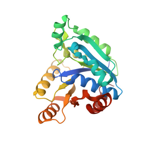
Precipitant-ligand exchange technique reveals the ADP binding mode in Mycobacterium tuberculosis dethiobiotin synthetase.

Thompson, A.P.Wegener, K.L.Booker, G.W.Polyak, S.W.Bruning, J.B.

(2018) Acta Crystallogr D Struct Biol 74: 965-972

  • DOI: https://doi.org/10.1107/S2059798318010136
  • Primary Citation of Related Structures:  
    6CZD, 6E05, 6E06

  • PubMed Abstract: 

    Dethiobiotin synthetase from Mycobacterium tuberculosis (MtDTBS) is a promising antituberculosis drug target. Small-molecule inhibitors that target MtDTBS provide a route towards new therapeutics for the treatment of antibiotic-resistant tuberculosis. Adenosine diphosphate (ADP) is an inhibitor of MtDTBS; however, structural studies into its mechanism of inhibition have been unsuccessful owing to competitive binding to the enzyme by crystallographic precipitants such as citrate and sulfate. Here, a crystallographic technique termed precipitant-ligand exchange has been developed to exchange protein-bound precipitants with ligands of interest. Proof of concept for the exchange method was demonstrated using cytidine triphosphate (CTP), which adopted the same binding mechanism as that obtained with traditional crystal-soaking techniques. Precipitant-ligand exchange also yielded the previously intractable structure of MtDTBS in complex with ADP solved to 2.4 Å resolution. This result demonstrates the utility of precipitant-ligand exchange, which may be widely applicable to protein crystallography.


  • Organizational Affiliation

    Molecular and Biomedical Science, The University of Adelaide, North Terrace, Adelaide, South Australia 5005, Australia.


Macromolecules
Find similar proteins by:  (by identity cutoff)  |  3D Structure
Entity ID: 1
MoleculeChains Sequence LengthOrganismDetailsImage
ATP-dependent dethiobiotin synthetase BioD
A, B, C, D
235Mycobacterium tuberculosis H37RvMutation(s): 0 
Gene Names: bioDRv1570MTCY336.33c
EC: 6.3.3.3
UniProt
Find proteins for P9WPQ5 (Mycobacterium tuberculosis (strain ATCC 25618 / H37Rv))
Explore P9WPQ5 
Go to UniProtKB:  P9WPQ5
Entity Groups  
Sequence Clusters30% Identity50% Identity70% Identity90% Identity95% Identity100% Identity
UniProt GroupP9WPQ5
Sequence Annotations
Expand
  • Reference Sequence
Experimental Data & Validation

Experimental Data

  • Method: X-RAY DIFFRACTION
  • Resolution: 2.50 Å
  • R-Value Free: 0.288 
  • R-Value Work: 0.217 
  • R-Value Observed: 0.220 
  • Space Group: P 21 21 21
Unit Cell:
Length ( Å )Angle ( ˚ )
a = 56.68α = 90
b = 105.7β = 90
c = 153.26γ = 90
Software Package:
Software NamePurpose
MOSFLMdata reduction
Aimlessdata scaling
PHASERphasing
PHENIXrefinement
PDB_EXTRACTdata extraction

Structure Validation

View Full Validation Report


Ligand Structure Quality Assessment 


Entry History & Funding Information

Deposition Data


Funding OrganizationLocationGrant Number
National Health and Medical Research Council (NHMRC, Australia)AustraliaAPP1068885

Revision History  (Full details and data files)

  • Version 1.0: 2018-10-17
    Type: Initial release
  • Version 1.1: 2020-01-08
    Changes: Author supporting evidence
  • Version 1.2: 2023-10-11
    Changes: Data collection, Database references, Derived calculations, Refinement description