


Crystal structure of the D141N variant of catalase-peroxidase from B. pseudomallei
Loewen, P.C.To be published.
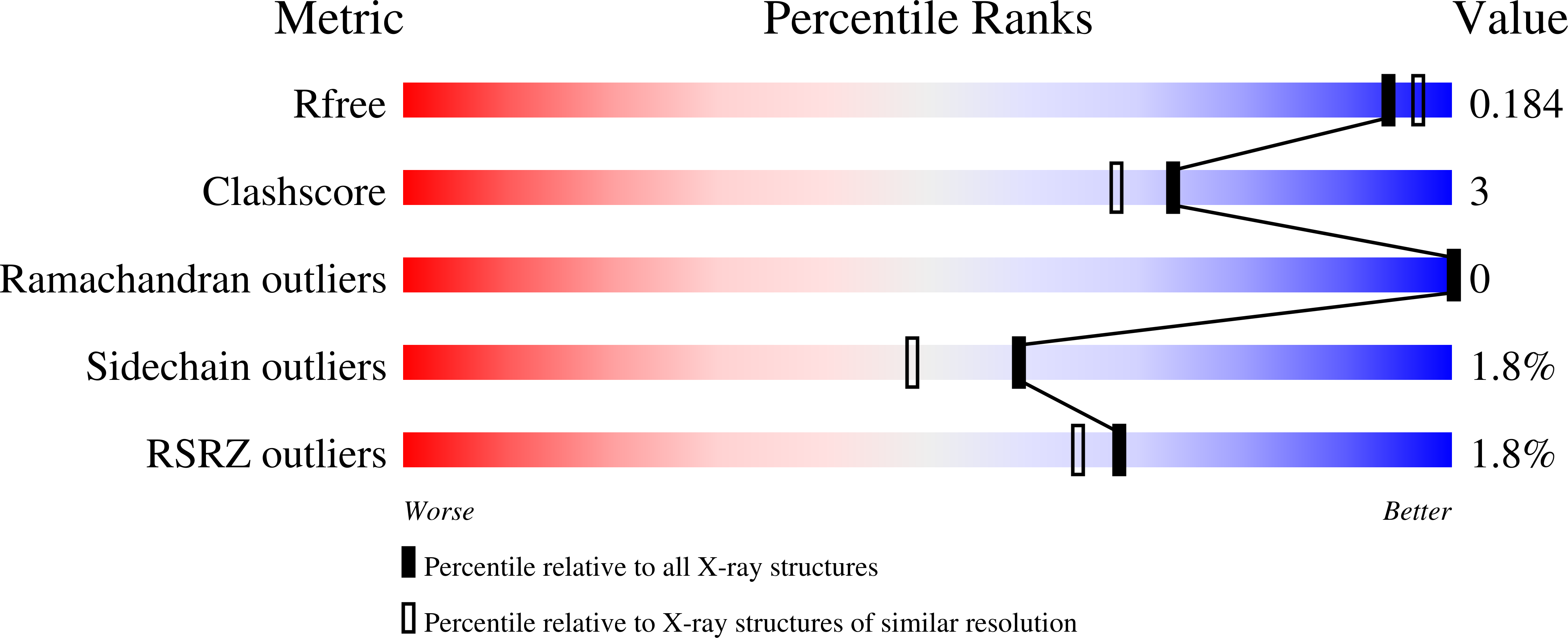
Experimental Data Snapshot
Entity ID: 1 | |||||
|---|---|---|---|---|---|
| Molecule | Chains | Sequence Length | Organism | Details | Image |
| Catalase-peroxidase | 728 | Burkholderia pseudomallei 1710b | Mutation(s): 1 Gene Names: katG, BURPS1710b_3366 EC: 1.11.1.21 | 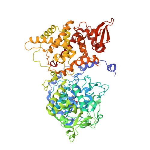 | |
UniProt | |||||
Find proteins for Q3JNW6 (Burkholderia pseudomallei (strain 1710b)) Explore Q3JNW6 Go to UniProtKB: Q3JNW6 | |||||
Entity Groups | |||||
| Sequence Clusters | 30% Identity50% Identity70% Identity90% Identity95% Identity100% Identity | ||||
| UniProt Group | Q3JNW6 | ||||
Sequence AnnotationsExpand | |||||
| |||||
| Ligands 6 Unique | |||||
|---|---|---|---|---|---|
| ID | Chains | Name / Formula / InChI Key | 2D Diagram | 3D Interactions | |
| HEM Query on HEM | C [auth A], K [auth B] | PROTOPORPHYRIN IX CONTAINING FE C34 H32 Fe N4 O4 KABFMIBPWCXCRK-RGGAHWMASA-L |  | ||
| MPD Query on MPD | H [auth A], I [auth A], J [auth A], P [auth B] | (4S)-2-METHYL-2,4-PENTANEDIOL C6 H14 O2 SVTBMSDMJJWYQN-YFKPBYRVSA-N |  | ||
| PO4 Query on PO4 | G [auth A], O [auth B] | PHOSPHATE ION O4 P NBIIXXVUZAFLBC-UHFFFAOYSA-K |  | ||
| CL Query on CL | E [auth A], M [auth B] | CHLORIDE ION Cl VEXZGXHMUGYJMC-UHFFFAOYSA-M |  | ||
| OXY Query on OXY | F [auth A], N [auth B] | OXYGEN MOLECULE O2 MYMOFIZGZYHOMD-UHFFFAOYSA-N |  | ||
| NA Query on NA | D [auth A], L [auth B] | SODIUM ION Na FKNQFGJONOIPTF-UHFFFAOYSA-N |  | ||
| Modified Residues 1 Unique | |||||
|---|---|---|---|---|---|
| ID | Chains | Type | Formula | 2D Diagram | Parent |
| TOX Query on TOX | A, B | L-PEPTIDE LINKING | C11 H12 N2 O4 |  | TRP |
| Length ( Å ) | Angle ( ˚ ) |
|---|---|
| a = 100.896 | α = 90 |
| b = 113.257 | β = 90 |
| c = 174.598 | γ = 90 |
| Software Name | Purpose |
|---|---|
| SCALA | data scaling |
| REFMAC | refinement |
| PDB_EXTRACT | data extraction |
| XDS | data reduction |
| MOLREP | phasing |
| Funding Organization | Location | Grant Number |
|---|---|---|
| Natural Sciences and Engineering Research Council (NSERC, Canada) | Canada | D9600-2012 |
| Canada Research Chair Program | Canada | -- |
RCSB PDB (citation) is hosted by
RCSB PDB is a member of the

