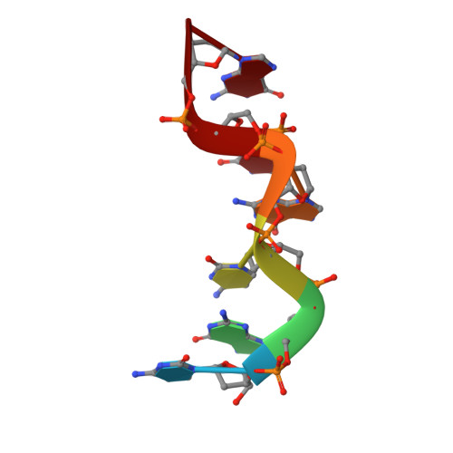
Four highly pseudosymmetric and/or twinned structures of d(CGCGCG)2 extend the repertoire of crystal structures of Z-DNA.
Luo, Z., Dauter, Z., Gilski, M.(2017) Acta Crystallogr D Struct Biol 73: 940-951
- PubMed: 29095165
- DOI: https://doi.org/10.1107/S2059798317014954
- Primary Citation of Related Structures:
6AQT, 6AQV, 6AQW, 6AQX - PubMed Abstract:
DNA oligomer duplexes containing alternating cytosines and guanines in their sequences tend to form left-handed helices of the Z-DNA type, with the sugar and phosphate backbone in a zigzag conformation and a helical repeat of two successive nucleotides. Z-DNA duplexes usually crystallize as hexagonally arranged parallel helical tubes, with various relative orientations and translation of neighboring duplexes. Four novel high-resolution crystal structures of d(CGCGCG) 2 duplexes are described here. They are characterized by a high degree of pseudosymmetry and/or twinning, with three or four independent duplexes differently oriented in a monoclinic P2 1 lattice of hexagonal metric. The various twinning criteria give somewhat conflicting indications in these complicated cases of crystal pathology. The details of molecular packing in these crystal structures are compared with other known crystal forms of Z-DNA.
Organizational Affiliation:
Synchrotron Radiation Research Section, MCL, National Cancer Institute, Argonne National Laboratory, Argonne, IL 60439, USA.