6DC6

Crystal structure of human ubiquitin activating enzyme E1 (Uba1) in complex with ubiquitin


Experimental Data Snapshot

  • Method: X-RAY DIFFRACTION
  • Resolution: 3.14 Å
  • R-Value Free: 0.250 
  • R-Value Work: 0.212 
  • R-Value Observed: 0.214 

wwPDB Validation   3D Report Full Report


This is version 1.4 of the entry. See complete history


Literature

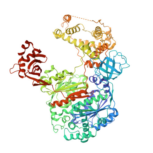
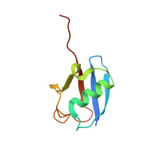
Crystal structure of a human ubiquitin E1-ubiquitin complex reveals conserved functional elements essential for activity.

Lv, Z.Williams, K.M.Yuan, L.Atkison, J.H.Olsen, S.K.

(2018) J Biol Chem 293: 18337-18352

  • DOI: https://doi.org/10.1074/jbc.RA118.003975
  • Primary Citation of Related Structures:  
    6DC6

  • PubMed Abstract: 

    Ubiquitin (Ub) signaling plays a key regulatory role in nearly every aspect of eukaryotic biology and is initiated by E1 enzymes that activate and transfer Ub to E2 Ub-conjugating enzymes. Despite Ub E1's fundamental importance to the cell and its attractiveness as a target for therapeutic intervention in cancer and other diseases, its only available structural information is derived from yeast orthologs of human ubiquitin-like modifier-activating enzyme 1 (hUBA1). To illuminate structural differences between yeast and hUBA1 structures that might be exploited for the development of small-molecule therapeutics, we determined the first crystal structure of a hUBA1-Ub complex. Using structural analysis, molecular modeling, and biochemical analysis, we demonstrate that hUBA1 shares a conserved overall structure and mechanism with previously characterized yeast orthologs, but displays subtle structural differences, particularly within the active site. Computational analysis revealed four potential ligand-binding hot spots on the surface of hUBA1 that might serve as targets to inhibit hUBA1 at the level of Ub activation or E2 recruitment or that might potentially be used in approaches such as protein-targeting chimeric molecules. Taken together, our work enhances our understanding of the hUBA1 mechanism, provides an improved framework for the development of small-molecule inhibitors of UBA1, and serves as a stepping stone for structural studies that involve the enzymes of the human Ub system at the level of both E1 and E2.


  • Organizational Affiliation

    From the Department of Biochemistry and Molecular Biology and Hollings Cancer Center, Medical University of South Carolina, Charleston, South Carolina 29425.


Macromolecules
Find similar proteins by:  (by identity cutoff)  |  3D Structure
Entity ID: 1
MoleculeChains Sequence LengthOrganismDetailsImage
Ubiquitin-like modifier-activating enzyme 1
A, C
1,015Homo sapiensMutation(s): 1 
Gene Names: UBA1A1S9TUBE1
EC: 6.2.1.45
UniProt & NIH Common Fund Data Resources
Find proteins for P22314 (Homo sapiens)
Explore P22314 
Go to UniProtKB:  P22314
PHAROS:  P22314
GTEx:  ENSG00000130985 
Entity Groups  
Sequence Clusters30% Identity50% Identity70% Identity90% Identity95% Identity100% Identity
UniProt GroupP22314
Sequence Annotations
Expand
  • Reference Sequence
Find similar proteins by:  (by identity cutoff)  |  3D Structure
Entity ID: 2
MoleculeChains Sequence LengthOrganismDetailsImage
Ubiquitin
B, D
80Homo sapiensMutation(s): 0 
UniProt & NIH Common Fund Data Resources
Find proteins for P62979 (Homo sapiens)
Explore P62979 
Go to UniProtKB:  P62979
PHAROS:  P62979
GTEx:  ENSG00000143947 
Entity Groups  
Sequence Clusters30% Identity50% Identity70% Identity90% Identity95% Identity100% Identity
UniProt GroupP62979
Sequence Annotations
Expand
  • Reference Sequence
Experimental Data & Validation

Experimental Data

  • Method: X-RAY DIFFRACTION
  • Resolution: 3.14 Å
  • R-Value Free: 0.250 
  • R-Value Work: 0.212 
  • R-Value Observed: 0.214 
  • Space Group: P 1 21 1
Unit Cell:
Length ( Å )Angle ( ˚ )
a = 104.999α = 90
b = 70.438β = 95.19
c = 188.851γ = 90
Software Package:
Software NamePurpose
PHENIXrefinement
HKL-2000data reduction
HKL-2000data scaling
PDB_EXTRACTdata extraction
PHASERphasing

Structure Validation

View Full Validation Report


Entry History & Funding Information

Deposition Data


Funding OrganizationLocationGrant Number
National Institutes of Health/National Institute of General Medical Sciences (NIH/NIGMS)United StatesR01GM115568

Revision History  (Full details and data files)

  • Version 1.0: 2018-10-10
    Type: Initial release
  • Version 1.1: 2018-10-17
    Changes: Data collection, Database references, Structure summary
  • Version 1.2: 2018-12-05
    Changes: Data collection, Database references
  • Version 1.3: 2020-01-01
    Changes: Author supporting evidence
  • Version 1.4: 2023-10-11
    Changes: Data collection, Database references, Derived calculations, Refinement description