5WBG

Crystal Structure of human Cytochrome P450 2B6 (Y226H/K262R) in complex with an analog of a drug Efavirenz

  • Classification: OXIDOREDUCTASE
  • Organism(s): Homo sapiens
  • Expression System: Escherichia coli
  • Mutation(s): Yes 

  • Deposited: 2017-06-29 Released: 2018-05-02 
  • Deposition Author(s): Shah, M.B., Halpert, J.R.
  • Funding Organization(s): National Institutes of Health/National Institute of Environmental Health Sciences (NIH/NIEHS)

Experimental Data Snapshot

  • Method: X-RAY DIFFRACTION
  • Resolution: 2.99 Å
  • R-Value Free: 0.262 
  • R-Value Work: 0.227 
  • R-Value Observed: 0.229 

wwPDB Validation   3D Report Full Report


Ligand Structure Quality Assessment 


This is version 1.2 of the entry. See complete history


Literature

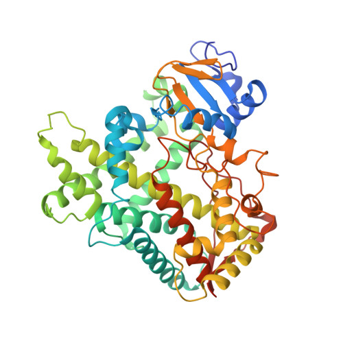
Crystal Structure of CYP2B6 in Complex with an Efavirenz Analog.

Shah, M.B.Zhang, Q.Halpert, J.R.

(2018) Int J Mol Sci 19

  • DOI: https://doi.org/10.3390/ijms19041025
  • Primary Citation of Related Structures:  
    5WBG

  • PubMed Abstract: 

    The over two dozen CYP2B structures of human, rabbit, and woodrat enzymes solved in the last decade have significantly enhanced our understanding of the structure-function relationships of drug metabolizing enzymes. More recently, an important role has emerged for halogen-π interactions in the CYP2B6 active site in substrate selectivity, explaining in part the preference for halogenated ligands as substrates. The mechanism by which such ligands interact with CYP2B enzymes involves conserved phenylalanine side chains, in particular F108, F115, or F297, in the active site, which form π bonds with halogens. To illustrate such halogen-π interactions using drugs that are major substrates of CYP2B6, we present here a crystal structure of CYP2B6 in complex with an analog of the widely used anti-HIV drug efavirenz, which contains a methyl group in place of the carbonyl oxygen. The chlorine of the efavirenz analog forms a π bond with the aromatic ring of F108, whereas the putative metabolism site on the distal end of the molecule is oriented towards the heme iron. The crystal structure showcases how CYP2B6 accommodates this important drug analog of considerable size in the active site by movement of various side chains without substantially increasing the active site volume. Furthermore, the CYP2B6-efavirenz analog complex provides a useful platform to investigate computationally as well as biophysically the effect of genetic polymorphisms on binding of the widely studied efavirenz.


  • Organizational Affiliation

    School of Pharmacy, University of Connecticut, Storrs, CT 06269, USA. manish.shah@acphs.edu.


Macromolecules
Find similar proteins by:  (by identity cutoff)  |  3D Structure
Entity ID: 1
MoleculeChains Sequence LengthOrganismDetailsImage
Cytochrome P450 2B6
A, B, C, D, E
A, B, C, D, E, F
476Homo sapiensMutation(s): 11 
Gene Names: CYP2B6
EC: 1.14.13
UniProt & NIH Common Fund Data Resources
Find proteins for P20813 (Homo sapiens)
Explore P20813 
Go to UniProtKB:  P20813
PHAROS:  P20813
GTEx:  ENSG00000197408 
Entity Groups  
Sequence Clusters30% Identity50% Identity70% Identity90% Identity95% Identity100% Identity
UniProt GroupP20813
Sequence Annotations
Expand
  • Reference Sequence
Small Molecules
Ligands 4 Unique
IDChains Name / Formula / InChI Key2D Diagram3D Interactions
HEM
Query on HEM

Download Ideal Coordinates CCD File 
G [auth A]
J [auth B]
M [auth C]
P [auth D]
S [auth E]
G [auth A],
J [auth B],
M [auth C],
P [auth D],
S [auth E],
V [auth F]
PROTOPORPHYRIN IX CONTAINING FE
C34 H32 Fe N4 O4
KABFMIBPWCXCRK-RGGAHWMASA-L
CM5
Query on CM5

Download Ideal Coordinates CCD File 
L [auth B],
O [auth C],
R [auth D],
U [auth E],
W [auth F]
5-CYCLOHEXYL-1-PENTYL-BETA-D-MALTOSIDE
C23 H42 O11
RVTGFZGNOSKUDA-ZNGNCRBCSA-N
9ZJ
Query on 9ZJ

Download Ideal Coordinates CCD File 
H [auth A]
K [auth B]
N [auth C]
Q [auth D]
T [auth E]
H [auth A],
K [auth B],
N [auth C],
Q [auth D],
T [auth E],
X [auth F]
(2R,4S)-6-chloro-4-(cyclopropylethynyl)-2-methyl-4-(trifluoromethyl)-1,4-dihydro-2H-3,1-benzoxazine
C15 H13 Cl F3 N O
IMPMOBJZWFZYGR-OTYXRUKQSA-N
ZAZ
Query on ZAZ

Download Ideal Coordinates CCD File 
I [auth A]5-cyclohexylpentan-1-ol
C11 H22 O
DOUCFSUZXLHVHJ-UHFFFAOYSA-N
Experimental Data & Validation

Experimental Data

  • Method: X-RAY DIFFRACTION
  • Resolution: 2.99 Å
  • R-Value Free: 0.262 
  • R-Value Work: 0.227 
  • R-Value Observed: 0.229 
  • Space Group: P 1 21 1
Unit Cell:
Length ( Å )Angle ( ˚ )
a = 103.293α = 90
b = 197.786β = 98.51
c = 119.222γ = 90
Software Package:
Software NamePurpose
REFMACrefinement
XDSdata reduction
SCALAdata scaling
PHASERphasing

Structure Validation

View Full Validation Report


Ligand Structure Quality Assessment 


Entry History & Funding Information

Deposition Data


Funding OrganizationLocationGrant Number
National Institutes of Health/National Institute of Environmental Health Sciences (NIH/NIEHS)United StatesES003619

Revision History  (Full details and data files)

  • Version 1.0: 2018-05-02
    Type: Initial release
  • Version 1.1: 2019-12-11
    Changes: Author supporting evidence
  • Version 1.2: 2023-10-04
    Changes: Data collection, Database references, Refinement description