5ONO

Crystal Structure of Ectoine Synthase from P. lautus


Experimental Data Snapshot

  • Method: X-RAY DIFFRACTION
  • Resolution: 2.50 Å
  • R-Value Free: 0.240 
  • R-Value Work: 0.185 
  • R-Value Observed: 0.191 

wwPDB Validation   3D Report Full Report


This is version 1.3 of the entry. See complete history


Literature

Illuminating the catalytic core of ectoine synthase through structural and biochemical analysis.

Czech, L.Hoppner, A.Kobus, S.Seubert, A.Riclea, R.Dickschat, J.S.Heider, J.Smits, S.H.J.Bremer, E.

(2019) Sci Rep 9: 364-364

  • DOI: https://doi.org/10.1038/s41598-018-36247-w
  • Primary Citation of Related Structures:  
    5ONM, 5ONN, 5ONO

  • PubMed Abstract: 

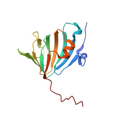
    Ectoine synthase (EctC) is the signature enzyme for the production of ectoine, a compatible solute and chemical chaperone widely synthesized by bacteria as a cellular defense against the detrimental effects of osmotic stress. EctC catalyzes the last step in ectoine synthesis through cyclo-condensation of the EctA-formed substrate N-gamma-acetyl-L-2,4-diaminobutyric acid via a water elimination reaction. We have biochemically and structurally characterized the EctC enzyme from the thermo-tolerant bacterium Paenibacillus lautus (Pl). EctC is a member of the cupin superfamily and forms dimers, both in solution and in crystals. We obtained high-resolution crystal structures of the (Pl)EctC protein in forms that contain (i) the catalytically important iron, (ii) iron and the substrate N-gamma-acetyl-L-2,4-diaminobutyric acid, and (iii) iron and the enzyme reaction product ectoine. These crystal structures lay the framework for a proposal for the EctC-mediated water-elimination reaction mechanism. Residues involved in coordinating the metal, the substrate, or the product within the active site of ectoine synthase are highly conserved among a large group of EctC-type proteins. Collectively, the biochemical, mutational, and structural data reported here yielded detailed insight into the structure-function relationship of the (Pl)EctC enzyme and are relevant for a deeper understanding of the ectoine synthase family as a whole.


  • Organizational Affiliation

    Department of Biology, Laboratory for Microbiology, Philipps-University Marburg, D-35043, Marburg, Germany.


Macromolecules
Find similar proteins by:  (by identity cutoff)  |  3D Structure
Entity ID: 1
MoleculeChains Sequence LengthOrganismDetailsImage
L-ectoine synthase138Paenibacillus lautusMutation(s): 0 
Gene Names: ectCBK123_26285
EC: 4.2.1.108
UniProt
Find proteins for A0A2A5LBI6 (Paenibacillus lautus)
Explore A0A2A5LBI6 
Go to UniProtKB:  A0A2A5LBI6
Entity Groups  
Sequence Clusters30% Identity50% Identity70% Identity90% Identity95% Identity100% Identity
UniProt GroupA0A2A5LBI6
Sequence Annotations
Expand
  • Reference Sequence
Small Molecules
Ligands 2 Unique
IDChains Name / Formula / InChI Key2D Diagram3D Interactions
4CS
Query on 4CS

Download Ideal Coordinates CCD File 
C [auth A](4S)-2-METHYL-1,4,5,6-TETRAHYDROPYRIMIDINE-4-CARBOXYLIC ACID
C6 H10 N2 O2
WQXNXVUDBPYKBA-YFKPBYRVSA-N
FE
Query on FE

Download Ideal Coordinates CCD File 
B [auth A]FE (III) ION
Fe
VTLYFUHAOXGGBS-UHFFFAOYSA-N
Experimental Data & Validation

Experimental Data

  • Method: X-RAY DIFFRACTION
  • Resolution: 2.50 Å
  • R-Value Free: 0.240 
  • R-Value Work: 0.185 
  • R-Value Observed: 0.191 
  • Space Group: P 31 2 1
Unit Cell:
Length ( Å )Angle ( ˚ )
a = 71.418α = 90
b = 71.418β = 90
c = 68.838γ = 120
Software Package:
Software NamePurpose
PHENIXrefinement
XDSdata reduction
XSCALEdata scaling
PHASERphasing

Structure Validation

View Full Validation Report


Entry History 

Deposition Data

  • Released Date: 2018-08-22 
  • Deposition Author(s): Bremer, E.

Revision History  (Full details and data files)

  • Version 1.0: 2018-08-22
    Type: Initial release
  • Version 1.1: 2019-03-06
    Changes: Data collection, Database references
  • Version 1.2: 2019-10-16
    Changes: Data collection
  • Version 1.3: 2024-01-17
    Changes: Data collection, Database references, Refinement description