5NJI

Structure of the dehydratase domain of PpsC from Mycobacterium tuberculosis in complex with C12:1-CoA


Experimental Data Snapshot

  • Method: X-RAY DIFFRACTION
  • Resolution: 1.60 Å
  • R-Value Free: 0.192 
  • R-Value Work: 0.170 
  • R-Value Observed: 0.171 

wwPDB Validation   3D Report Full Report


Ligand Structure Quality Assessment 


This is version 1.2 of the entry. See complete history


Literature

Insights into Substrate Modification by Dehydratases from Type I Polyketide Synthases.

Faille, A.Gavalda, S.Slama, N.Lherbet, C.Maveyraud, L.Guillet, V.Laval, F.Quemard, A.Mourey, L.Pedelacq, J.D.

(2017) J Mol Biol 429: 1554-1569

  • DOI: https://doi.org/10.1016/j.jmb.2017.03.026
  • Primary Citation of Related Structures:  
    5I0K, 5L84, 5NJI

  • PubMed Abstract: 

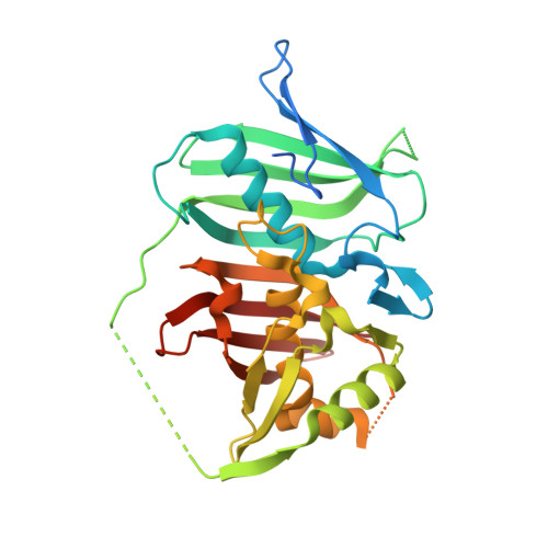
    Dehydration reactions play a crucial role in the de novo biosynthesis of fatty acids and a wide range of pharmacologically active polyketide natural products with strong emphasis on human medicine. The type I polyketide synthase PpsC from Mycobacterium tuberculosis catalyzes key biosynthetic steps of lipid virulence factors phthiocerol dimycocerosates and phenolic glycolipids. Given the insolubility of the natural C 28 -C 30 fatty acyl substrate of the PpsC dehydratase (DH) domain, we investigated its structure-function relationships in the presence of shorter surrogate substrates. Since most enzymes belonging to the (R)-specific enoyl hydratase/hydroxyacyl dehydratase family conduct the reverse hydration reaction in vitro, we have determined the X-ray structures of the PpsC DH domain, both unliganded (apo) and in complex with trans-but-2-enoyl-CoA or trans-dodec-2-enoyl-CoA derivatives. This study provides for the first time a snapshot of dehydratase-ligand interactions following a hydration reaction. Our structural analysis allowed us to identify residues essential for substrate binding and activity. The structural comparison of the two complexes also sheds light on the need for long acyl chains for this dehydratase to carry out its function, consistent with both its in vitro catalytic behavior and the physiological role of the PpsC enzyme.


  • Organizational Affiliation

    Institut de Pharmacologie et de Biologie Structurale, Université de Toulouse, CNRS, UPS, 31077 Toulouse Cedex 04, France.


Macromolecules
Find similar proteins by:  (by identity cutoff)  |  3D Structure
Entity ID: 1
MoleculeChains Sequence LengthOrganismDetailsImage
Phthiocerol/phenolphthiocerol synthesis polyketide synthase type I PpsC322Mycobacterium tuberculosis H37RvMutation(s): 1 
Gene Names: ppsCRv2933
EC: 2.3.1.41
UniProt
Find proteins for P96202 (Mycobacterium tuberculosis (strain ATCC 25618 / H37Rv))
Explore P96202 
Go to UniProtKB:  P96202
Entity Groups  
Sequence Clusters30% Identity50% Identity70% Identity90% Identity95% Identity100% Identity
UniProt GroupP96202
Sequence Annotations
Expand
  • Reference Sequence
Small Molecules
Ligands 1 Unique
IDChains Name / Formula / InChI Key2D Diagram3D Interactions
8Z2
Query on 8Z2

Download Ideal Coordinates CCD File 
B [auth A]~{S}-[2-[3-[[(2~{R})-4-[[[(2~{R},3~{S},4~{R},5~{R})-5-(6-aminopurin-9-yl)-4-oxidanyl-3-phosphonooxy-oxolan-2-yl]methoxy-oxidanyl-phosphoryl]oxy-oxidanyl-phosphoryl]oxy-3,3-dimethyl-2-oxidanyl-butanoyl]amino]propanoylamino]ethyl] (~{E})-dodec-2-enethioate
C33 H56 N7 O17 P3 S
IRFYVBULXZMEDE-DEEZISNZSA-N
Experimental Data & Validation

Experimental Data

  • Method: X-RAY DIFFRACTION
  • Resolution: 1.60 Å
  • R-Value Free: 0.192 
  • R-Value Work: 0.170 
  • R-Value Observed: 0.171 
  • Space Group: C 1 2 1
Unit Cell:
Length ( Å )Angle ( ˚ )
a = 117.377α = 90
b = 70.81β = 104.87
c = 42.571γ = 90
Software Package:
Software NamePurpose
PHENIXrefinement
XDSdata reduction
Cootmodel building
PHENIXphasing

Structure Validation

View Full Validation Report


Ligand Structure Quality Assessment 


Entry History 

Deposition Data

Revision History  (Full details and data files)

  • Version 1.0: 2017-05-31
    Type: Initial release
  • Version 1.1: 2017-06-07
    Changes: Database references
  • Version 1.2: 2024-01-17
    Changes: Data collection, Database references, Refinement description