5M4O

Crystal structure of hydroquinone 1,2-dioxygenase from Sphingomonas sp. TTNP3 in complex with 4-nitrophenol


Experimental Data Snapshot

  • Method: X-RAY DIFFRACTION
  • Resolution: 2.10 Å
  • R-Value Free: 0.249 
  • R-Value Work: 0.188 
  • R-Value Observed: 0.191 

wwPDB Validation   3D Report Full Report


This is version 1.0 of the entry. See complete history


Literature

The crystal structures of native hydroquinone 1,2-dioxygenase from Sphingomonas sp. TTNP3 and of substrate and inhibitor complexes.

Ferraroni, M.Da Vela, S.Kolvenbach, B.A.Corvini, P.F.Scozzafava, A.

(2017) Biochim Biophys Acta 1865: 520-530

  • DOI: https://doi.org/10.1016/j.bbapap.2017.02.013
  • Primary Citation of Related Structures:  
    5M21, 5M22, 5M26, 5M4O

  • PubMed Abstract: 

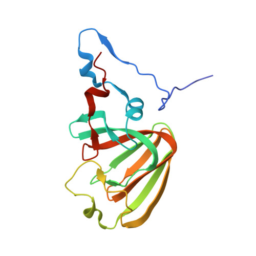
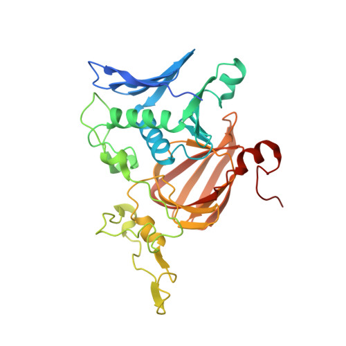
    The crystal structure of hydroquinone 1,2-dioxygenase, a Fe(II) ring cleaving dioxygenase from Sphingomonas sp. strain TTNP3, which oxidizes a wide range of hydroquinones to the corresponding 4-hydroxymuconic semialdehydes, has been solved by Molecular Replacement, using the coordinates of PnpCD from Pseudomonas sp. strain WBC-3. The enzyme is a heterotetramer, constituted of two subunits α and two β of 19 and 38kDa, respectively. Both the two subunits fold as a cupin, but that of the small α subunit lacks a competent metal binding pocket. Two tetramers are present in the asymmetric unit. Each of the four β subunits in the asymmetric unit binds one Fe(II) ion. The iron ion in each β subunit is coordinated to three protein residues, His258, Glu264, and His305 and a water molecule. The crystal structures of the complexes with the substrate methylhydroquinone, obtained under anaerobic conditions, and with the inhibitors 4-hydroxybenzoate and 4-nitrophenol were also solved. The structures of the native enzyme and of the complexes present significant differences in the active site region compared to PnpCD, the other hydroquinone 1,2-dioxygenase of known structure, and in particular they show a different coordination at the metal center.


  • Organizational Affiliation

    Dipartimento di Chimica "Ugo Schiff", Università di Firenze, Via della Lastruccia 3, I-50019, Sesto Fiorentino, FI, Italy. Electronic address: marta.ferraroni@unifi.it.


Macromolecules
Find similar proteins by:  (by identity cutoff)  |  3D Structure
Entity ID: 1
MoleculeChains Sequence LengthOrganismDetailsImage
Hydroquinone dioxygenase small subunit
A, C, E, G
170Sphingomonas sp. TTNP3Mutation(s): 0 
EC: 1.13.11
UniProt
Find proteins for F8TW82 (Sphingomonas sp. TTNP3)
Explore F8TW82 
Go to UniProtKB:  F8TW82
Entity Groups  
Sequence Clusters30% Identity50% Identity70% Identity90% Identity95% Identity100% Identity
UniProt GroupF8TW82
Sequence Annotations
Expand
  • Reference Sequence
Find similar proteins by:  (by identity cutoff)  |  3D Structure
Entity ID: 2
MoleculeChains Sequence LengthOrganismDetailsImage
Hydroquinone dioxygenase large subunit
B, D, F, H
341Sphingomonas sp. TTNP3Mutation(s): 0 
EC: 1.13.11
UniProt
Find proteins for F8TW83 (Sphingomonas sp. TTNP3)
Explore F8TW83 
Go to UniProtKB:  F8TW83
Entity Groups  
Sequence Clusters30% Identity50% Identity70% Identity90% Identity95% Identity100% Identity
UniProt GroupF8TW83
Sequence Annotations
Expand
  • Reference Sequence
Experimental Data & Validation

Experimental Data

  • Method: X-RAY DIFFRACTION
  • Resolution: 2.10 Å
  • R-Value Free: 0.249 
  • R-Value Work: 0.188 
  • R-Value Observed: 0.191 
  • Space Group: P 1 21 1
Unit Cell:
Length ( Å )Angle ( ˚ )
a = 88.992α = 90
b = 125.982β = 105.14
c = 92.331γ = 90
Software Package:
Software NamePurpose
XSCALEdata scaling
REFMACrefinement
PDB_EXTRACTdata extraction
XSCALEdata reduction
REFMACphasing

Structure Validation

View Full Validation Report


Entry History 

Deposition Data

Revision History  (Full details and data files)

  • Version 1.0: 2017-09-27
    Type: Initial release