


5KY2
mouse POFUT1 in complex with O-glucosylated mouse Factor VII EGF1 and GDP
- PDB DOI: https://doi.org/10.2210/pdb5KY2/pdb
- Classification: TRANSFERASE
- Organism(s): Mus musculus
- Expression System: Homo sapiens, Escherichia coli
- Mutation(s): No
- Deposited: 2016-07-20 Released: 2017-05-17
- Funding Organization(s): Canadian Institutes of Health Research (CIHR)
Experimental Data Snapshot
- Method: X-RAY DIFFRACTION
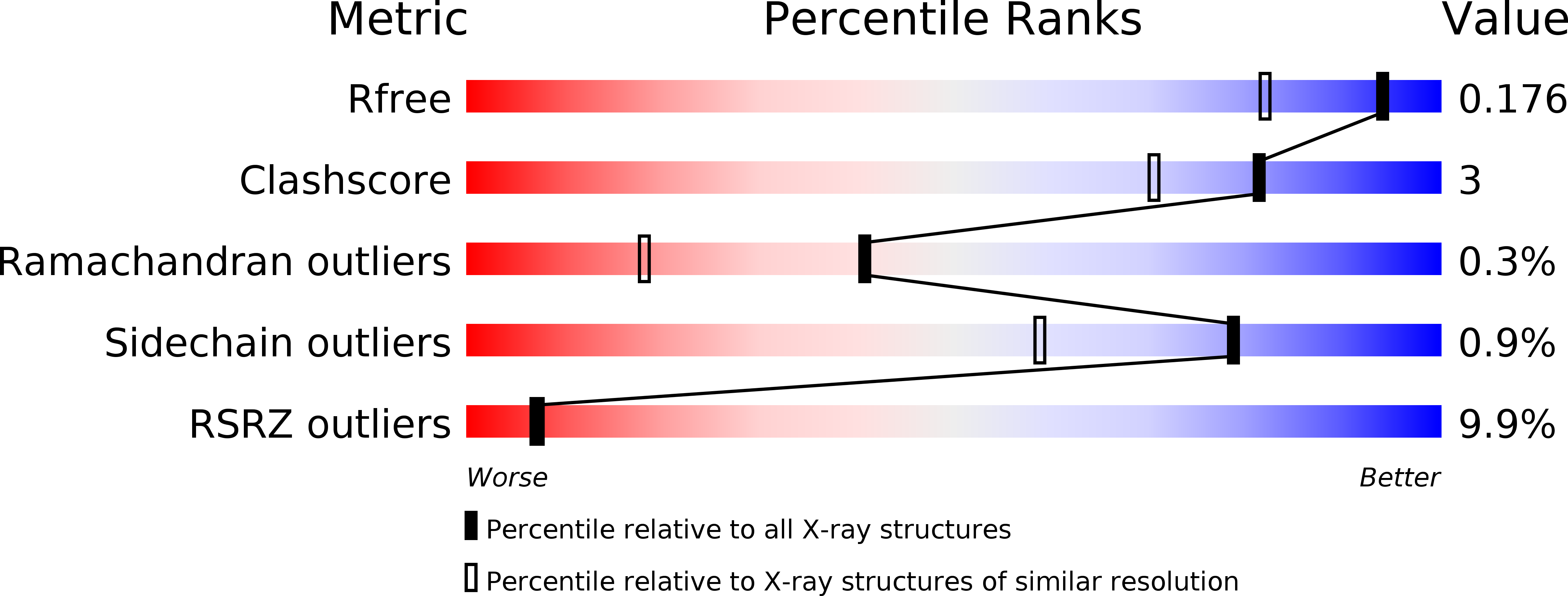
- Resolution: 1.47 Å
- R-Value Free: 0.175
- R-Value Work: 0.153
- R-Value Observed: 0.154
This is version 2.0 of the entry. See complete history.

Macromolecules
Find similar proteins by:
(by identity cutoff) | 3D Structure
Entity ID: 1 | |||||
|---|---|---|---|---|---|
| Molecule | Chains | Sequence Length | Organism | Details | Image |
| GDP-fucose protein O-fucosyltransferase 1 | 355 | Mus musculus | Mutation(s): 0 Gene Names: Pofut1 EC: 2.4.1.221 | 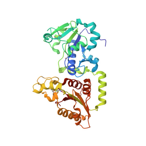 | |
UniProt | |||||
Find proteins for Q91ZW2 (Mus musculus) Explore Q91ZW2 Go to UniProtKB: Q91ZW2 | |||||
Entity Groups | |||||
| Sequence Clusters | 30% Identity50% Identity70% Identity90% Identity95% Identity100% Identity | ||||
| UniProt Group | Q91ZW2 | ||||
Sequence AnnotationsExpand | |||||
| |||||
Find similar proteins by:
(by identity cutoff) | 3D Structure
Entity ID: 2 | |||||
|---|---|---|---|---|---|
| Molecule | Chains | Sequence Length | Organism | Details | Image |
| Coagulation factor VII | 40 | Mus musculus | Mutation(s): 0 Gene Names: F7, Cf7 EC: 3.4.21.21 | 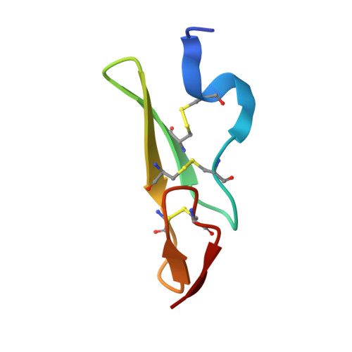 | |
UniProt & NIH Common Fund Data Resources | |||||
Find proteins for P70375 (Mus musculus) Explore P70375 Go to UniProtKB: P70375 | |||||
IMPC: MGI:109325 | |||||
Entity Groups | |||||
| Sequence Clusters | 30% Identity50% Identity70% Identity90% Identity95% Identity100% Identity | ||||
| UniProt Group | P70375 | ||||
Sequence AnnotationsExpand | |||||
| |||||
Small Molecules
| Ligands 4 Unique | |||||
|---|---|---|---|---|---|
| ID | Chains | Name / Formula / InChI Key | 2D Diagram | 3D Interactions | |
| GDP Query on GDP | E [auth A] | GUANOSINE-5'-DIPHOSPHATE C10 H15 N5 O11 P2 QGWNDRXFNXRZMB-UUOKFMHZSA-N |  | ||
| NAG Query on NAG | C [auth A], D [auth A] | 2-acetamido-2-deoxy-beta-D-glucopyranose C8 H15 N O6 OVRNDRQMDRJTHS-FMDGEEDCSA-N |  | ||
| BGC Query on BGC | I [auth B] | beta-D-glucopyranose C6 H12 O6 WQZGKKKJIJFFOK-VFUOTHLCSA-N |  | ||
| GOL Query on GOL | F [auth A], G [auth A], H [auth A] | GLYCEROL C3 H8 O3 PEDCQBHIVMGVHV-UHFFFAOYSA-N |  | ||
Experimental Data & Validation
Experimental Data
- Method: X-RAY DIFFRACTION
- Resolution: 1.47 Å
- R-Value Free: 0.175
- R-Value Work: 0.153
- R-Value Observed: 0.154
- Space Group: P 21 21 21
Unit Cell:
| Length ( Å ) | Angle ( ˚ ) |
|---|---|
| a = 51.965 | α = 90 |
| b = 67.138 | β = 90 |
| c = 109.357 | γ = 90 |
| Software Name | Purpose |
|---|---|
| XSCALE | data scaling |
| PHENIX | refinement |
| PDB_EXTRACT | data extraction |
| XDS | data reduction |
| PHASER | phasing |
Entry History & Funding Information
Deposition Data
- Released Date: 2017-05-17 Deposition Author(s): Li, Z., Rini, J.M.
| Funding Organization | Location | Grant Number |
|---|---|---|
| Canadian Institutes of Health Research (CIHR) | Canada | -- |
Revision History (Full details and data files)
- Version 1.0: 2017-05-17
Type: Initial release - Version 1.1: 2017-05-31
Changes: Database references - Version 1.2: 2017-06-28
Changes: Database references - Version 1.3: 2017-09-20
Changes: Author supporting evidence - Version 1.4: 2020-01-08
Changes: Author supporting evidence, Data collection - Version 2.0: 2020-07-29
Type: Remediation
Reason: Carbohydrate remediation
Changes: Atomic model, Data collection, Derived calculations, Structure summary
