5J1W

Crystal structure of human CLK1 in complex with pyrido[3,4-g]quinazoline derivative ZW31 (compound 14)


Experimental Data Snapshot

  • Method: X-RAY DIFFRACTION
  • Resolution: 2.42 Å
  • R-Value Free: 0.228 
  • R-Value Work: 0.189 
  • R-Value Observed: 0.191 

wwPDB Validation   3D Report Full Report


Ligand Structure Quality Assessment 


This is version 1.1 of the entry. See complete history


Literature

Discovery of pyrido[3,4-g]quinazoline derivatives as CMGC family protein kinase inhibitors: Design, synthesis, inhibitory potency and X-ray co-crystal structure.

Esvan, Y.J.Zeinyeh, W.Boibessot, T.Nauton, L.Thery, V.Knapp, S.Chaikuad, A.Loaec, N.Meijer, L.Anizon, F.Giraud, F.Moreau, P.

(2016) Eur J Med Chem 118: 170-177

  • DOI: https://doi.org/10.1016/j.ejmech.2016.04.004
  • Primary Citation of Related Structures:  
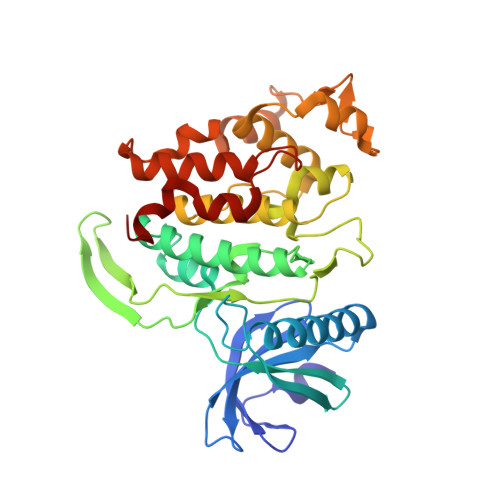
    5J1V, 5J1W

  • PubMed Abstract: 

    The design and synthesis of new pyrido[3,4-g]quinazoline derivatives is described as well as their protein kinase inhibitory potencies toward five CMGC family members (CDK5, CK1, GSK3, CLK1 and DYRK1A). The interest for this original tricyclic heteroaromatic scaffold as modulators of CLK1/DYRK1A activity was validated by nanomolar potencies (compounds 12 and 13). CLK1 co-crystal structures with two inhibitors revealed the binding mode of these compounds within the ATP-binding pocket.


  • Organizational Affiliation

    Université Clermont Auvergne, Université Blaise Pascal, Institut de Chimie de Clermont-Ferrand, BP 10448, F-63000 Clermont-Ferrand, France; CNRS, UMR 6296, ICCF, F-63178 Aubière, France.


Macromolecules
Find similar proteins by:  (by identity cutoff)  |  3D Structure
Entity ID: 1
MoleculeChains Sequence LengthOrganismDetailsImage
Dual specificity protein kinase CLK1
A, B, C
339Homo sapiensMutation(s): 0 
Gene Names: CLK1CLK
EC: 2.7.12.1
UniProt & NIH Common Fund Data Resources
Find proteins for P49759 (Homo sapiens)
Explore P49759 
Go to UniProtKB:  P49759
PHAROS:  P49759
GTEx:  ENSG00000013441 
Entity Groups  
Sequence Clusters30% Identity50% Identity70% Identity90% Identity95% Identity100% Identity
UniProt GroupP49759
Sequence Annotations
Expand
  • Reference Sequence
Small Molecules
Ligands 3 Unique
IDChains Name / Formula / InChI Key2D Diagram3D Interactions
6FB
Query on 6FB

Download Ideal Coordinates CCD File 
I [auth A],
L [auth B],
Q [auth C],
R [auth C]
pyrido[3,4-g]quinazolin-2-amine
C11 H8 N4
HTMYPIWEXZOFDM-UHFFFAOYSA-N
PO4
Query on PO4

Download Ideal Coordinates CCD File 
K [auth B],
P [auth C]
PHOSPHATE ION
O4 P
NBIIXXVUZAFLBC-UHFFFAOYSA-K
GOL
Query on GOL

Download Ideal Coordinates CCD File 
D [auth A]
E [auth A]
F [auth A]
G [auth A]
H [auth A]
D [auth A],
E [auth A],
F [auth A],
G [auth A],
H [auth A],
J [auth B],
M [auth C],
N [auth C],
O [auth C]
GLYCEROL
C3 H8 O3
PEDCQBHIVMGVHV-UHFFFAOYSA-N
Binding Affinity Annotations 
IDSourceBinding Affinity
6FB Binding MOAD:  5J1W IC50: 190 (nM) from 1 assay(s)
BindingDB:  5J1W IC50: 190 (nM) from 1 assay(s)
Experimental Data & Validation

Experimental Data

  • Method: X-RAY DIFFRACTION
  • Resolution: 2.42 Å
  • R-Value Free: 0.228 
  • R-Value Work: 0.189 
  • R-Value Observed: 0.191 
  • Space Group: P 1 21 1
Unit Cell:
Length ( Å )Angle ( ˚ )
a = 56.63α = 90
b = 118.04β = 99.55
c = 90.5γ = 90
Software Package:
Software NamePurpose
REFMACrefinement
iMOSFLMdata reduction
SCALAdata scaling
PHASERphasing

Structure Validation

View Full Validation Report


Ligand Structure Quality Assessment 


Entry History 

Revision History  (Full details and data files)

  • Version 1.0: 2016-05-04
    Type: Initial release
  • Version 1.1: 2016-05-11
    Changes: Database references