5HXW

L-amino acid deaminase from Proteus vulgaris


Experimental Data Snapshot

  • Method: X-RAY DIFFRACTION
  • Resolution: 2.63 Å
  • R-Value Free: 0.252 
  • R-Value Work: 0.221 
  • R-Value Observed: 0.223 

wwPDB Validation   3D Report Full Report


Ligand Structure Quality Assessment 


This is version 1.2 of the entry. See complete history


Literature

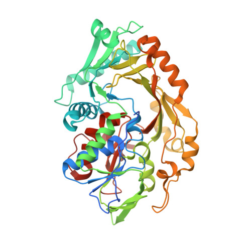
Crystal structure of a membrane-bound l-amino acid deaminase from Proteus vulgaris

Ju, Y.Tong, S.Gao, Y.Zhao, W.Liu, Q.Gu, Q.Xu, J.Niu, L.Teng, M.Zhou, H.

(2016) J Struct Biol 195: 306-315

  • DOI: https://doi.org/10.1016/j.jsb.2016.07.008
  • Primary Citation of Related Structures:  
    5HXW, 5I39

  • PubMed Abstract: 

    l-amino acid oxidases/deaminases (LAAOs/LAADs) are a class of oxidoreductases catalyzing the oxidative deamination of l-amino acids to α-keto acids. They are widely distributed in eukaryotic and prokaryotic organisms, and exhibit diverse substrate specificity, post-translational modifications and cellular localization. While LAAOs isolated from snake venom have been extensively characterized, the structures and functions of LAAOs from other species are largely unknown. Here, we reported crystal structure of a bacterial membrane-bound LAAD from Proteus vulgaris (pvLAAD) in complex with flavin adenine dinucleotide (FAD). We found that the overall fold of pvLAAD does not resemble typical LAAOs. Instead it, is similar to d-amino acid oxidases (DAAOs) with an additional hydrophobic insertion module on protein surface. Structural analysis and liposome-binding assays suggested that the hydrophobic module serves as an extra membrane-binding site for LAADs. Bacteria from genera Proteus and Providencia were found to encode two classes of membrane-bound LAADs. Based on our structure, the key roles of residues Q278 and L317 in substrate selectivity were proposed and biochemically analyzed. While LAADs on the membrane were proposed to transfer electrons to respiratory chain for FAD re-oxidization, we observed that the purified pvLAAD could generate a significant amount of hydrogen peroxide in vitro, suggesting it could use dioxygen to directly re-oxidize FADH2 as what typical LAAOs usually do. These findings provide a novel insights for a better understanding this class of enzymes and will help developing biocatalysts for industrial applications.


  • Organizational Affiliation

    Research Center for Structural Biology and Drug Discovery, School of Pharmaceutical Sciences, Sun Yat-sen University, Guangzhou 510006, China.


Macromolecules
Find similar proteins by:  (by identity cutoff)  |  3D Structure
Entity ID: 1
MoleculeChains Sequence LengthOrganismDetailsImage
L-amino acid deaminase
A, B, C, D, E
A, B, C, D, E, F
454Proteus vulgarisMutation(s): 0 
Gene Names: LAD
EC: 1.4.3.2
UniProt
Find proteins for Q9LCB2 (Proteus vulgaris)
Explore Q9LCB2 
Go to UniProtKB:  Q9LCB2
Entity Groups  
Sequence Clusters30% Identity50% Identity70% Identity90% Identity95% Identity100% Identity
UniProt GroupQ9LCB2
Sequence Annotations
Expand
  • Reference Sequence
Small Molecules
Ligands 2 Unique
IDChains Name / Formula / InChI Key2D Diagram3D Interactions
FAD
Query on FAD

Download Ideal Coordinates CCD File 
G [auth A]
K [auth B]
Q [auth C]
S [auth D]
W [auth E]
G [auth A],
K [auth B],
Q [auth C],
S [auth D],
W [auth E],
Y [auth F]
FLAVIN-ADENINE DINUCLEOTIDE
C27 H33 N9 O15 P2
VWWQXMAJTJZDQX-UYBVJOGSSA-N
16A
Query on 16A

Download Ideal Coordinates CCD File 
AA [auth F]
H [auth A]
I [auth A]
J [auth A]
L [auth B]
AA [auth F],
H [auth A],
I [auth A],
J [auth A],
L [auth B],
M [auth B],
N [auth B],
O [auth B],
P [auth B],
R [auth C],
T [auth D],
U [auth D],
V [auth D],
X [auth E],
Z [auth F]
CETYL-TRIMETHYL-AMMONIUM
C19 H42 N
RLGQACBPNDBWTB-UHFFFAOYSA-N
Experimental Data & Validation

Experimental Data

  • Method: X-RAY DIFFRACTION
  • Resolution: 2.63 Å
  • R-Value Free: 0.252 
  • R-Value Work: 0.221 
  • R-Value Observed: 0.223 
  • Space Group: P 1
Unit Cell:
Length ( Å )Angle ( ˚ )
a = 100.314α = 64.52
b = 104.575β = 73.06
c = 105.419γ = 61.17
Software Package:
Software NamePurpose
REFMACrefinement
HKL-2000data reduction
HKL-2000data scaling
MOLREPphasing

Structure Validation

View Full Validation Report


Ligand Structure Quality Assessment 


Entry History 

Deposition Data

Revision History  (Full details and data files)

  • Version 1.0: 2016-08-03
    Type: Initial release
  • Version 1.1: 2016-08-24
    Changes: Database references
  • Version 1.2: 2023-11-08
    Changes: Data collection, Database references, Derived calculations, Refinement description