5V39

Crystal structure of human vitamin D receptor ligand binding domain in complex with a VDRM


Experimental Data Snapshot

  • Method: X-RAY DIFFRACTION
  • Resolution: 2.20 Å
  • R-Value Free: 0.235 
  • R-Value Work: 0.167 
  • R-Value Observed: 0.172 

wwPDB Validation   3D Report Full Report


Ligand Structure Quality Assessment 


This is version 1.2 of the entry. See complete history


Literature

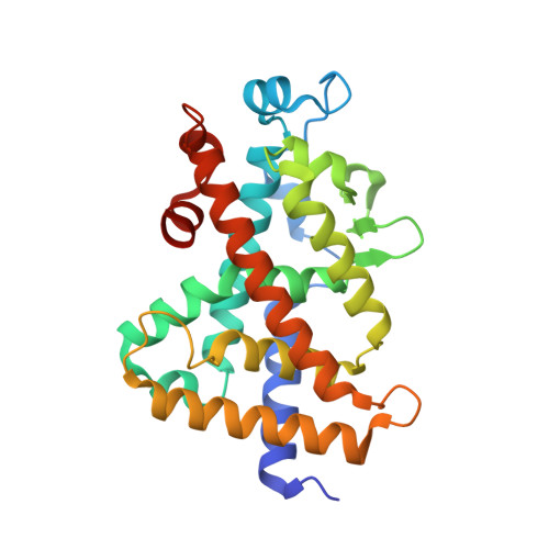
HDX reveals the conformational dynamics of DNA sequence specific VDR co-activator interactions.

Zheng, J.Chang, M.R.Stites, R.E.Wang, Y.Bruning, J.B.Pascal, B.D.Novick, S.J.Garcia-Ordonez, R.D.Stayrook, K.R.Chalmers, M.J.Dodge, J.A.Griffin, P.R.

(2017) Nat Commun 8: 923-923

  • DOI: https://doi.org/10.1038/s41467-017-00978-7
  • Primary Citation of Related Structures:  
    5V39

  • PubMed Abstract: 

    The vitamin D receptor/retinoid X receptor-α heterodimer (VDRRXRα) regulates bone mineralization via transcriptional control of osteocalcin (BGLAP) gene and is the receptor for 1α,25-dihydroxyvitamin D 3 (1,25D3). However, supra-physiological levels of 1,25D3 activates the calcium-regulating gene TRPV6 leading to hypercalcemia. An approach to attenuate this adverse effect is to develop selective VDR modulators (VDRMs) that differentially activate BGLAP but not TRPV6. Here we present structural insight for the action of a VDRM compared with agonists by employing hydrogen/deuterium exchange. Agonist binding directs crosstalk between co-receptors upon DNA binding, stabilizing the activation function 2 (AF2) surfaces of both receptors driving steroid receptor co-activator-1 (SRC1) interaction. In contrast, AF2 of VDR within VDRM:BGLAP bound heterodimer is more vulnerable for large stabilization upon SRC1 interaction compared with VDRM:TRPV6 bound heterodimer. These results reveal that the combination of ligand structure and DNA sequence tailor the transcriptional activity of VDR toward specific target genes.The vitamin D receptor/retinoid X receptor-α heterodimer (VDRRXRα) regulates bone mineralization. Here the authors employ hydrogen/deuterium exchange (HDX) mass spectrometry to study the conformational dynamics of VDRRXRα and give mechanistic insights into how VDRRXRα controls the transcriptional activity of specific genes.


  • Organizational Affiliation

    Department of Molecular Medicine, The Scripps Research Institute, Jupiter, FL, 33458, USA.


Macromolecules
Find similar proteins by:  (by identity cutoff)  |  3D Structure
Entity ID: 1
MoleculeChains Sequence LengthOrganismDetailsImage
Vitamin D3 receptor257Homo sapiensMutation(s): 0 
Gene Names: VDRNR1I1
UniProt & NIH Common Fund Data Resources
Find proteins for P11473 (Homo sapiens)
Explore P11473 
Go to UniProtKB:  P11473
PHAROS:  P11473
GTEx:  ENSG00000111424 
Entity Groups  
Sequence Clusters30% Identity50% Identity70% Identity90% Identity95% Identity100% Identity
UniProt GroupP11473
Sequence Annotations
Expand
  • Reference Sequence
Small Molecules
Ligands 1 Unique
IDChains Name / Formula / InChI Key2D Diagram3D Interactions
8VM
Query on 8VM

Download Ideal Coordinates CCD File 
B [auth A]5-(3-{4-[(2S)-2-hydroxy-3,3-dimethylbutoxy]-3-methylphenyl}pentan-3-yl)-3-methyl-N-(1H-tetrazol-5-yl)thiophene-2-carboxamide
C25 H35 N5 O3 S
XZOVDHKWBOZRPS-LJQANCHMSA-N
Experimental Data & Validation

Experimental Data

  • Method: X-RAY DIFFRACTION
  • Resolution: 2.20 Å
  • R-Value Free: 0.235 
  • R-Value Work: 0.167 
  • R-Value Observed: 0.172 
  • Space Group: P 21 21 21
Unit Cell:
Length ( Å )Angle ( ˚ )
a = 44α = 90
b = 52.67β = 90
c = 105.88γ = 90
Software Package:
Software NamePurpose
BUSTERrefinement
MOSFLMdata reduction
SCALAdata scaling
PHASERphasing

Structure Validation

View Full Validation Report


Ligand Structure Quality Assessment 


Entry History 

Deposition Data

Revision History  (Full details and data files)

  • Version 1.0: 2018-03-14
    Type: Initial release
  • Version 1.1: 2019-04-03
    Changes: Data collection, Database references
  • Version 1.2: 2024-03-06
    Changes: Data collection, Database references