5UT6

JAK2 JH2 in complex with a diaminopyrimidine


Experimental Data Snapshot

  • Method: X-RAY DIFFRACTION
  • Resolution: 1.65 Å
  • R-Value Free: 0.206 
  • R-Value Work: 0.166 
  • R-Value Observed: 0.169 

wwPDB Validation   3D Report Full Report


Ligand Structure Quality Assessment 


This is version 1.4 of the entry. See complete history


Literature

JAK2 JH2 Fluorescence Polarization Assay and Crystal Structures for Complexes with Three Small Molecules.

Newton, A.S.Deiana, L.Puleo, D.E.Cisneros, J.A.Cutrona, K.J.Schlessinger, J.Jorgensen, W.L.

(2017) ACS Med Chem Lett 8: 614-617

  • DOI: https://doi.org/10.1021/acsmedchemlett.7b00154
  • Primary Citation of Related Structures:  
    5UT4, 5UT5, 5UT6

  • PubMed Abstract: 

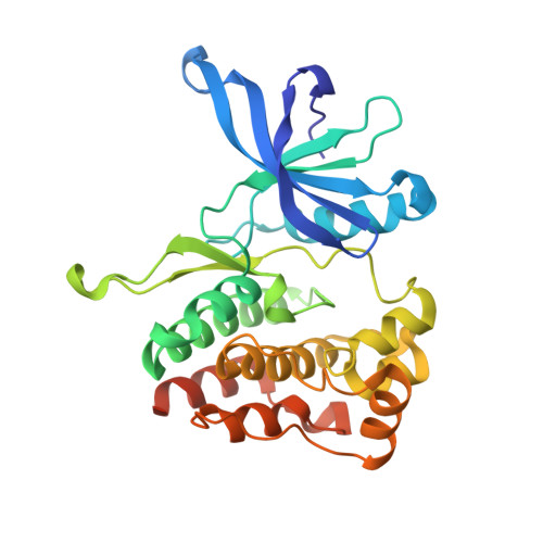
    A competitive fluorescence polarization (FP) assay is reported for determining binding affinities of probe molecules with the pseudokinase JAK2 JH2 allosteric site. The syntheses of the fluorescent 5 and 6 used in the assay are reported as well as K d results for 10 compounds, including JNJ7706621, NVP-BSK805, and filgotinib (GLPG0634). X-ray crystal structures of JAK2 JH2 in complex with NVP-BSK805, filgotinib, and diaminopyrimidine 8 elucidate the binding poses.


  • Organizational Affiliation

    Department of Chemistry, Yale University, New Haven, Connecticut 06520-8107, United States.


Macromolecules
Find similar proteins by:  (by identity cutoff)  |  3D Structure
Entity ID: 1
MoleculeChains Sequence LengthOrganismDetailsImage
Tyrosine-protein kinase JAK2289Homo sapiensMutation(s): 3 
Gene Names: JAK2
EC: 2.7.10.2
UniProt & NIH Common Fund Data Resources
Find proteins for O60674 (Homo sapiens)
Explore O60674 
Go to UniProtKB:  O60674
PHAROS:  O60674
GTEx:  ENSG00000096968 
Entity Groups  
Sequence Clusters30% Identity50% Identity70% Identity90% Identity95% Identity100% Identity
UniProt GroupO60674
Sequence Annotations
Expand
  • Reference Sequence
Small Molecules
Ligands 4 Unique
IDChains Name / Formula / InChI Key2D Diagram3D Interactions
8MY
Query on 8MY

Download Ideal Coordinates CCD File 
B [auth A]4-({4-amino-6-[3-(hydroxymethyl)-1H-pyrazol-1-yl]pyrimidin-2-yl}amino)benzonitrile
C15 H13 N7 O
NMBKNACBCUDEER-UHFFFAOYSA-N
GOL
Query on GOL

Download Ideal Coordinates CCD File 
C [auth A],
D [auth A],
E [auth A],
F [auth A],
G [auth A]
GLYCEROL
C3 H8 O3
PEDCQBHIVMGVHV-UHFFFAOYSA-N
DMS
Query on DMS

Download Ideal Coordinates CCD File 
H [auth A]DIMETHYL SULFOXIDE
C2 H6 O S
IAZDPXIOMUYVGZ-UHFFFAOYSA-N
ACT
Query on ACT

Download Ideal Coordinates CCD File 
I [auth A]ACETATE ION
C2 H3 O2
QTBSBXVTEAMEQO-UHFFFAOYSA-M
Binding Affinity Annotations 
IDSourceBinding Affinity
8MY BindingDB:  5UT6 Kd: 5.73e+4 (nM) from 1 assay(s)
Binding MOAD:  5UT6 Kd: 5.73e+4 (nM) from 1 assay(s)
Experimental Data & Validation

Experimental Data

  • Method: X-RAY DIFFRACTION
  • Resolution: 1.65 Å
  • R-Value Free: 0.206 
  • R-Value Work: 0.166 
  • R-Value Observed: 0.169 
  • Space Group: P 1 21 1
Unit Cell:
Length ( Å )Angle ( ˚ )
a = 44.371α = 90
b = 57.642β = 110.24
c = 60.682γ = 90
Software Package:
Software NamePurpose
PHENIXrefinement
XDSdata reduction
XDSdata scaling
PHASERphasing

Structure Validation

View Full Validation Report


Ligand Structure Quality Assessment 


Entry History & Funding Information

Deposition Data


Funding OrganizationLocationGrant Number
National Institutes of Health/National Institute of General Medical Sciences (NIH/NIGMS)United StatesGM007324
Gilead SciencesUnited StatesYG-003-13

Revision History  (Full details and data files)

  • Version 1.0: 2017-06-07
    Type: Initial release
  • Version 1.1: 2017-07-05
    Changes: Database references
  • Version 1.2: 2017-09-13
    Changes: Author supporting evidence
  • Version 1.3: 2020-01-01
    Changes: Author supporting evidence
  • Version 1.4: 2023-10-04
    Changes: Data collection, Database references, Refinement description