5U2S

Pre-catalytic ternary complex of Human DNA Polymerase Beta With Gapped DNA substrate incoming (-)3TC-TP and Ca2+.


Experimental Data Snapshot

  • Method: X-RAY DIFFRACTION
  • Resolution: 2.30 Å
  • R-Value Free: 0.284 
  • R-Value Work: 0.230 
  • R-Value Observed: 0.233 

wwPDB Validation   3D Report Full Report


Ligand Structure Quality Assessment 


This is version 1.5 of the entry. See complete history


Literature

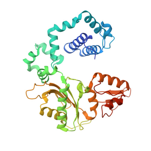
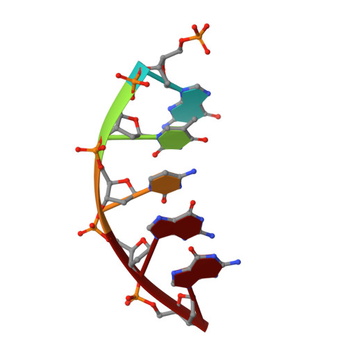
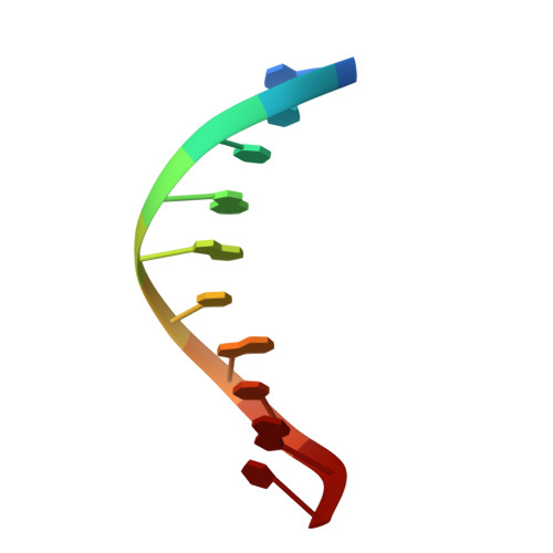
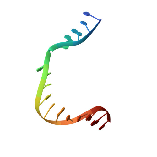
Structural basis for the D-stereoselectivity of human DNA polymerase beta.

Vyas, R.Reed, A.J.Raper, A.T.Zahurancik, W.J.Wallenmeyer, P.C.Suo, Z.

(2017) Nucleic Acids Res 45: 6228-6237

  • DOI: https://doi.org/10.1093/nar/gkx252
  • Primary Citation of Related Structures:  
    5U2R, 5U2S, 5U2T

  • PubMed Abstract: 

    Nucleoside reverse transcriptase inhibitors (NRTIs) with L-stereochemistry have long been an effective treatment for viral infections because of the strong D-stereoselectivity exhibited by human DNA polymerases relative to viral reverse transcriptases. The D-stereoselectivity of DNA polymerases has only recently been explored structurally and all three DNA polymerases studied to date have demonstrated unique stereochemical selection mechanisms. Here, we have solved structures of human DNA polymerase β (hPolβ), in complex with single-nucleotide gapped DNA and L-nucleotides and performed pre-steady-state kinetic analysis to determine the D-stereoselectivity mechanism of hPolβ. Beyond a similar 180° rotation of the L-nucleotide ribose ring seen in other studies, the pre-catalytic ternary crystal structures of hPolβ, DNA and L-dCTP or the triphosphate forms of antiviral drugs lamivudine ((-)3TC-TP) and emtricitabine ((-)FTC-TP) provide little structural evidence to suggest that hPolβ follows the previously characterized mechanisms of D-stereoselectivity. Instead, hPolβ discriminates against L-stereochemistry through accumulation of several active site rearrangements that lead to a decreased nucleotide binding affinity and incorporation rate. The two NRTIs escape some of the active site selection through the base and sugar modifications but are selected against through the inability of hPolβ to complete thumb domain closure.


  • Organizational Affiliation

    Department of Chemistry and Biochemistry, The Ohio State University, Columbus, OH 43210, USA.


Macromolecules

Find similar proteins by:  (by identity cutoff)  |  3D Structure
Entity ID: 1
MoleculeChains Sequence LengthOrganismDetailsImage
DNA polymerase beta343Homo sapiensMutation(s): 0 
Gene Names: POLB
EC: 2.7.7.7 (PDB Primary Data), 4.2.99 (PDB Primary Data)
UniProt & NIH Common Fund Data Resources
Find proteins for P06746 (Homo sapiens)
Explore P06746 
Go to UniProtKB:  P06746
PHAROS:  P06746
GTEx:  ENSG00000070501 
Entity Groups  
Sequence Clusters30% Identity50% Identity70% Identity90% Identity95% Identity100% Identity
UniProt GroupP06746
Sequence Annotations
Expand
  • Reference Sequence

Find similar nucleic acids by:  Sequence   |   3D Structure  

Entity ID: 2
MoleculeChains LengthOrganismImage
5-MER PHOSPHORYLATED DOWNSTREAM PRIMERB [auth D]5synthetic construct
Sequence Annotations
Expand
  • Reference Sequence

Find similar nucleic acids by:  Sequence   |   3D Structure  

Entity ID: 3
MoleculeChains LengthOrganismImage
10- MER PRIMERC [auth P]10synthetic construct
Sequence Annotations
Expand
  • Reference Sequence

Find similar nucleic acids by:  Sequence   |   3D Structure  

Entity ID: 4
MoleculeChains LengthOrganismImage
16- MER TEMPLATED [auth T]16synthetic construct
Sequence Annotations
Expand
  • Reference Sequence
Small Molecules
Ligands 4 Unique
IDChains Name / Formula / InChI Key2D Diagram3D Interactions
1RZ
Query on 1RZ

Download Ideal Coordinates CCD File 
E [auth A]Lamivudine Triphosphate
C8 H14 N3 O12 P3 S
YLEQMGZZMCJKCN-NKWVEPMBSA-N
ACT
Query on ACT

Download Ideal Coordinates CCD File 
J [auth A]
K [auth A]
L [auth A]
M [auth A]
N [auth A]
J [auth A],
K [auth A],
L [auth A],
M [auth A],
N [auth A],
O [auth A]
ACETATE ION
C2 H3 O2
QTBSBXVTEAMEQO-UHFFFAOYSA-M
CA
Query on CA

Download Ideal Coordinates CCD File 
F [auth A]CALCIUM ION
Ca
BHPQYMZQTOCNFJ-UHFFFAOYSA-N
NA
Query on NA

Download Ideal Coordinates CCD File 
G [auth A],
H [auth A],
I [auth A],
P [auth A]
SODIUM ION
Na
FKNQFGJONOIPTF-UHFFFAOYSA-N
Experimental Data & Validation

Experimental Data

  • Method: X-RAY DIFFRACTION
  • Resolution: 2.30 Å
  • R-Value Free: 0.284 
  • R-Value Work: 0.230 
  • R-Value Observed: 0.233 
  • Space Group: P 1 21 1
Unit Cell:
Length ( Å )Angle ( ˚ )
a = 54.63α = 90
b = 79.45β = 106.66
c = 55.17γ = 90
Software Package:
Software NamePurpose
MOSFLMdata reduction
SCALEPACKdata scaling
PHASERphasing
REFMACrefinement

Structure Validation

View Full Validation Report


Ligand Structure Quality Assessment 


Entry History & Funding Information

Deposition Data


Funding OrganizationLocationGrant Number
National Institutes of Health/National Institute of Environmental Health Sciences (NIH/NIEHS)United StatesES024585 and ES026821
National Science Foundation (NSF, United States)United StatesMCB-0960961

Revision History  (Full details and data files)

  • Version 1.0: 2017-05-24
    Type: Initial release
  • Version 1.1: 2017-06-14
    Changes: Database references
  • Version 1.2: 2017-09-13
    Changes: Author supporting evidence
  • Version 1.3: 2019-07-17
    Changes: Data collection, Derived calculations, Structure summary
  • Version 1.4: 2019-11-27
    Changes: Author supporting evidence
  • Version 1.5: 2023-10-04
    Changes: Data collection, Database references, Derived calculations, Refinement description