Structure of phosphorylated UBL domain and insights into PINK1-orchestrated parkin activation.
Aguirre, J.D., Dunkerley, K.M., Mercier, P., Shaw, G.S.(2017) Proc Natl Acad Sci U S A 114: 298-303
- PubMed: 28007983
- DOI: https://doi.org/10.1073/pnas.1613040114
- Primary Citation of Related Structures:
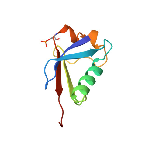
5TR5 - PubMed Abstract:
Mutations in PARK2 and PARK6 genes are responsible for the majority of hereditary Parkinson's disease cases. These genes encode the E3 ubiquitin ligase parkin and the protein kinase PTEN-induced kinase 1 (PINK1), respectively. Together, parkin and PINK1 regulate the mitophagy pathway, which recycles damaged mitochondria following oxidative stress. Native parkin is inactive and exists in an autoinhibited state mediated by its ubiquitin-like (UBL) domain. PINK1 phosphorylation of serine 65 in parkin's UBL and serine 65 of ubiquitin fully activate ubiquitin ligase activity; however, a structural rationale for these observations is not clear. Here, we report the structure of the phosphorylated UBL domain from parkin. We find that destabilization of the UBL results from rearrangements to hydrophobic core packing that modify its structure. Altered surface electrostatics from the phosphoserine group disrupt its intramolecular association, resulting in poorer autoinhibition in phosphorylated parkin. Further, we show that phosphorylation of both the UBL domain and ubiquitin are required to activate parkin by releasing the UBL domain, forming an extended structure needed to facilitate E2-ubiquitin binding. Together, the results underscore the importance of parkin activation by the PINK1 phosphorylation signal and provide a structural picture of the unraveling of parkin's ubiquitin ligase potential.
Organizational Affiliation:
Department of Biochemistry, Schulich School of Medicine & Dentistry, University of Western Ontario, London, ON N6A 3K7, Canada.