5SWK

Crystal structure of p53 epitope-scaffold based on a inhibitor of cysteine proteases in complex with human MDM2


Experimental Data Snapshot

  • Method: X-RAY DIFFRACTION
  • Resolution: 1.92 Å
  • R-Value Free: 0.249 
  • R-Value Work: 0.221 
  • R-Value Observed: 0.223 

wwPDB Validation   3D Report Full Report


This is version 1.3 of the entry. See complete history


Literature

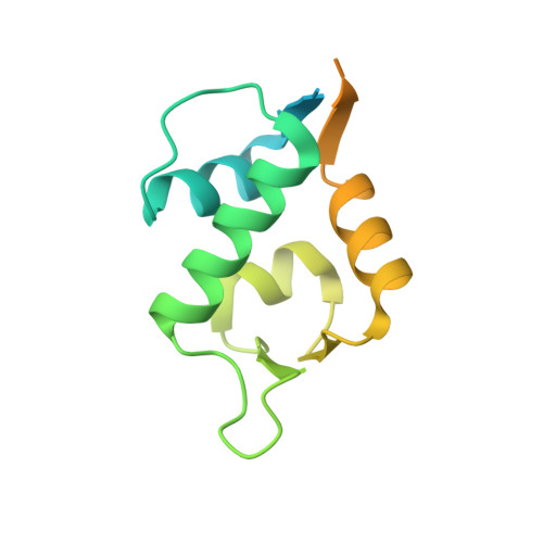
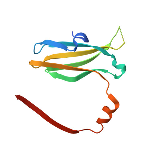
Mimicking a p53-MDM2 interaction based on a stable immunoglobulin-like domain scaffold.

Jimenez-Sandoval, P.Madrigal-Carrillo, E.A.Santamaria-Suarez, H.A.Maturana, D.Renteria-Gonzalez, I.Benitez-Cardoza, C.G.Torres-Larios, A.Brieba, L.G.

(2018) Proteins 86: 802-812

  • DOI: https://doi.org/10.1002/prot.25519
  • Primary Citation of Related Structures:  
    5SWK

  • PubMed Abstract: 

    Antibodies recognize protein targets with great affinity and specificity. However, posttranslational modifications and the presence of intrinsic disulfide-bonds pose difficulties for their industrial use. The immunoglobulin fold is one of the most ubiquitous folds in nature and it is found in many proteins besides antibodies. An example of a protein family with an immunoglobulin-like fold is the Cysteine Protease Inhibitors (ICP) family I42 of the MEROPs database for protease and protease inhibitors. Members of this protein family are thermostable and do not present internal disulfide bonds. Crystal structures of several ICPs indicate that they resemble the Ig-like domain of the human T cell co-receptor CD8α As ICPs present 2 flexible recognition loops that vary accordingly to their targeted protease, we hypothesize that members of this protein family would be ideal to design peptide aptamers that mimic protein-protein interactions. Herein, we use an ICP variant from Entamoeba histolytica (EhICP1) to mimic the interaction between p53 and MDM2. We found that a 13 amino-acid peptide derived from p53 can be introduced in 2 variable loops (DE, FG) but not the third (BC). Chimeric EhICP1-p53 form a stable complex with MDM2 at a micromolar range. Crystal structure of the EhICP1-p53(FG)-loop variant in complex with MDM2 reveals a swapping subdomain between 2 chimeric molecules, however, the p53 peptide interacts with MDM2 as in previous crystal structures. The structural details of the EhICP1-p53(FG) interaction with MDM2 resemble the interaction between an antibody and MDM2.


  • Organizational Affiliation

    Laboratorio Nacional de Genómica para la Biodiversidad, Centro de Investigación y de Estudios Avanzados del IPN, Apartado Postal 629, Irapuato, Guanajuato, CP 36821, México.


Macromolecules
Find similar proteins by:  (by identity cutoff)  |  3D Structure
Entity ID: 1
MoleculeChains Sequence LengthOrganismDetailsImage
E3 ubiquitin-protein ligase Mdm2
A, B
153Homo sapiensMutation(s): 0 
Gene Names: MDM2
EC: 6.3.2
UniProt & NIH Common Fund Data Resources
Find proteins for Q00987 (Homo sapiens)
Explore Q00987 
Go to UniProtKB:  Q00987
PHAROS:  Q00987
GTEx:  ENSG00000135679 
Entity Groups  
Sequence Clusters30% Identity50% Identity70% Identity90% Identity95% Identity100% Identity
UniProt GroupQ00987
Sequence Annotations
Expand
  • Reference Sequence
Find similar proteins by:  (by identity cutoff)  |  3D Structure
Entity ID: 2
MoleculeChains Sequence LengthOrganismDetailsImage
De novo protein based on the inhibitor Amoebiasin-1
C, D
110Entamoeba histolyticaMutation(s): 0 
Gene Names: AMSICP1
Entity Groups  
Sequence Clusters30% Identity50% Identity70% Identity90% Identity95% Identity100% Identity
Sequence Annotations
Expand
  • Reference Sequence
Small Molecules
Ligands 3 Unique
IDChains Name / Formula / InChI Key2D Diagram3D Interactions
SO4
Query on SO4

Download Ideal Coordinates CCD File 
G [auth B],
L [auth C]
SULFATE ION
O4 S
QAOWNCQODCNURD-UHFFFAOYSA-L
GOL
Query on GOL

Download Ideal Coordinates CCD File 
N [auth D]GLYCEROL
C3 H8 O3
PEDCQBHIVMGVHV-UHFFFAOYSA-N
CL
Query on CL

Download Ideal Coordinates CCD File 
E [auth A]
F [auth A]
H [auth B]
I [auth B]
J [auth B]
E [auth A],
F [auth A],
H [auth B],
I [auth B],
J [auth B],
K [auth B],
M [auth D]
CHLORIDE ION
Cl
VEXZGXHMUGYJMC-UHFFFAOYSA-M
Experimental Data & Validation

Experimental Data

  • Method: X-RAY DIFFRACTION
  • Resolution: 1.92 Å
  • R-Value Free: 0.249 
  • R-Value Work: 0.221 
  • R-Value Observed: 0.223 
  • Space Group: P 43 21 2
Unit Cell:
Length ( Å )Angle ( ˚ )
a = 87.361α = 90
b = 87.361β = 90
c = 160.84γ = 90
Software Package:
Software NamePurpose
Aimlessdata scaling
PHASERphasing
PHENIXrefinement
PDB_EXTRACTdata extraction
iMOSFLMdata processing
Cootmodel building
iMOSFLMdata reduction

Structure Validation

View Full Validation Report


Entry History & Funding Information

Deposition Data


Funding OrganizationLocationGrant Number
Consejo Nacional de Ciencia y Tecnologia (CONACYT)MexicoFronteras de la ciencia No. 11

Revision History  (Full details and data files)

  • Version 1.0: 2017-10-18
    Type: Initial release
  • Version 1.1: 2019-05-01
    Changes: Data collection, Database references
  • Version 1.2: 2020-01-15
    Changes: Author supporting evidence
  • Version 1.3: 2023-10-04
    Changes: Data collection, Database references, Refinement description