5O3J

Crystal structure of TIA-1 RRM2 in complex with RNA


Experimental Data Snapshot

  • Method: X-RAY DIFFRACTION
  • Resolution: 2.97 Å
  • R-Value Free: 0.312 
  • R-Value Work: 0.251 
  • R-Value Observed: 0.254 

wwPDB Validation   3D Report Full Report


This is version 1.3 of the entry. See complete history


Literature

Segmental, Domain-Selective Perdeuteration and Small-Angle Neutron Scattering for Structural Analysis of Multi-Domain Proteins.

Sonntag, M.Jagtap, P.K.A.Simon, B.Appavou, M.S.Geerlof, A.Stehle, R.Gabel, F.Hennig, J.Sattler, M.

(2017) Angew Chem Int Ed Engl 56: 9322-9325

  • DOI: https://doi.org/10.1002/anie.201702904
  • Primary Citation of Related Structures:  
    5O2V, 5O3J

  • PubMed Abstract: 

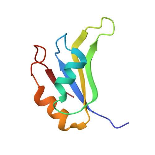
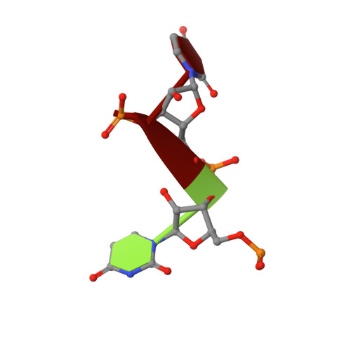
    Multi-domain proteins play critical roles in fine-tuning essential processes in cellular signaling and gene regulation. Typically, multiple globular domains that are connected by flexible linkers undergo dynamic rearrangements upon binding to protein, DNA or RNA ligands. RNA binding proteins (RBPs) represent an important class of multi-domain proteins, which regulate gene expression by recognizing linear or structured RNA sequence motifs. Here, we employ segmental perdeuteration of the three RNA recognition motif (RRM) domains in the RBP TIA-1 using Sortase A mediated protein ligation. We show that domain-selective perdeuteration combined with contrast-matched small-angle neutron scattering (SANS), SAXS and computational modeling provides valuable information to precisely define relative domain arrangements. The approach is generally applicable to study conformational arrangements of individual domains in multi-domain proteins and changes induced by ligand binding.


  • Organizational Affiliation

    Institute of Structural Biology, Helmholtz Zentrum München, Ingolstädter Landstr. 1, 85764, Neuherberg, Germany.


Macromolecules

Find similar proteins by:  (by identity cutoff)  |  3D Structure
Entity ID: 1
MoleculeChains Sequence LengthOrganismDetailsImage
Nucleolysin TIA-1 isoform p4080Homo sapiensMutation(s): 0 
Gene Names: TIA1
UniProt & NIH Common Fund Data Resources
Find proteins for P31483 (Homo sapiens)
Explore P31483 
Go to UniProtKB:  P31483
PHAROS:  P31483
GTEx:  ENSG00000116001 
Entity Groups  
Sequence Clusters30% Identity50% Identity70% Identity90% Identity95% Identity100% Identity
UniProt GroupP31483
Sequence Annotations
Expand
  • Reference Sequence

Find similar nucleic acids by:  Sequence   |   3D Structure  

Entity ID: 2
MoleculeChains LengthOrganismImage
RNA (5'-R(P*UP*UP*C)-3')3Homo sapiens
Sequence Annotations
Expand
  • Reference Sequence
Experimental Data & Validation

Experimental Data

  • Method: X-RAY DIFFRACTION
  • Resolution: 2.97 Å
  • R-Value Free: 0.312 
  • R-Value Work: 0.251 
  • R-Value Observed: 0.254 
  • Space Group: P 65
Unit Cell:
Length ( Å )Angle ( ˚ )
a = 44.31α = 90
b = 44.31β = 90
c = 85.72γ = 120
Software Package:
Software NamePurpose
REFMACrefinement
XDSdata reduction
XSCALEdata scaling
PHASERphasing

Structure Validation

View Full Validation Report


Entry History 

Deposition Data

Revision History  (Full details and data files)

  • Version 1.0: 2017-07-05
    Type: Initial release
  • Version 1.1: 2017-08-09
    Changes: Database references
  • Version 1.2: 2019-10-16
    Changes: Data collection
  • Version 1.3: 2024-01-17
    Changes: Data collection, Database references, Refinement description