


Insights into Gasdermin D activation from the crystal structure of its C-terminal domain
Anton, L., Sborgi, L., Hiller, S., Broz, P., Maier, T.(2017) Biorxiv
Experimental Data Snapshot
wwPDB Validation 3D Report Full Report

(2017) Biorxiv
Entity ID: 1 | |||||
|---|---|---|---|---|---|
| Molecule | Chains | Sequence Length | Organism | Details | Image |
| Gasdermin-D | 207 | Homo sapiens | Mutation(s): 0 Gene Names: GSDMD, DFNA5L, GSDMDC1, FKSG10 | 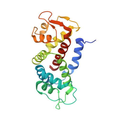 | |
UniProt & NIH Common Fund Data Resources | |||||
Find proteins for P57764 (Homo sapiens) Explore P57764 Go to UniProtKB: P57764 | |||||
PHAROS: P57764 GTEx: ENSG00000104518 | |||||
Entity Groups | |||||
| Sequence Clusters | 30% Identity50% Identity70% Identity90% Identity95% Identity100% Identity | ||||
| UniProt Group | P57764 | ||||
Sequence AnnotationsExpand | |||||
| |||||
| Length ( Å ) | Angle ( ˚ ) |
|---|---|
| a = 125.73 | α = 90 |
| b = 47.98 | β = 90 |
| c = 37.65 | γ = 90 |
| Software Name | Purpose |
|---|---|
| PHENIX | refinement |
| XDS | data reduction |
| XSCALE | data scaling |
| PHASER | phasing |
RCSB PDB (citation) is hosted by
RCSB PDB is a member of the

