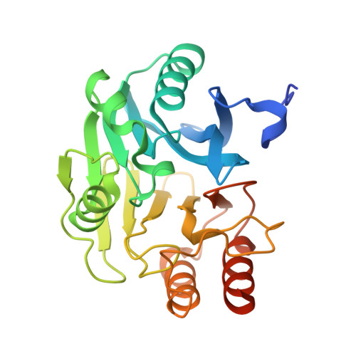
Crystal structures of VIM-1 complexes explain active site heterogeneity in VIM-class metallo-beta-lactamases.
Salimraj, R., Hinchliffe, P., Kosmopoulou, M., Tyrrell, J.M., Brem, J., van Berkel, S.S., Verma, A., Owens, R.J., McDonough, M.A., Walsh, T.R., Schofield, C.J., Spencer, J.(2019) FEBS J 286: 169-183
- PubMed: 30430727
- DOI: https://doi.org/10.1111/febs.14695
- Primary Citation of Related Structures:
5N5G, 5N5H, 5N5I - PubMed Abstract:
Metallo-β-Lactamases (MBLs) protect bacteria from almost all β-lactam antibiotics. Verona integron-encoded MBL (VIM) enzymes are among the most clinically important MBLs, with VIM-1 increasing in carbapenem-resistant Enterobacteriaceae (Escherichia coli, Klebsiella pneumoniae) that are among the hardest bacterial pathogens to treat. VIM enzymes display sequence variation at residues (224 and 228) that in related MBLs are conserved and participate in substrate binding. How they accommodate this variability, while retaining catalytic efficiency against a broad substrate range, has remained unclear. Here, we present crystal structures of VIM-1 and its complexes with a substrate-mimicking thioenolate inhibitor, ML302F, that restores meropenem activity against a range of VIM-1 producing clinical strains, and the hydrolysed product of the carbapenem meropenem. Comparison of these two structures identifies a water-mediated hydrogen bond, between the carboxylate group of substrate/inhibitor and the backbone carbonyl of the active site zinc ligand Cys221, that is common to both complexes. Structural comparisons show that the responsible Cys221-bound water is observed in all known VIM structures, participates in carboxylate binding with other inhibitor classes, and thus effectively replicates the role of the conserved Lys224 in analogous complexes with other MBLs. These results provide a mechanism for substrate binding that permits the variation at positions 224 and 228 that is a hallmark of VIM MBLs. ENZYMES: EC 3.5.2.6 DATABASES: Co-ordinates and structure factors for protein structures described in this manuscript have been deposited in the Protein Data Bank (www.rcsb.org/pdb) with accession codes 5N5G (VIM-1), 5N5H (VIM-1:ML302F complex) and 5N5I (VIM-1-hydrolysed meropenem complex).
Organizational Affiliation:
School of Cellular and Molecular Medicine, University of Bristol, UK.