5MY6

Crystal structure of a HER2-Nb complex


Experimental Data Snapshot

  • Method: X-RAY DIFFRACTION
  • Resolution: 2.25 Å
  • R-Value Free: 0.220 
  • R-Value Work: 0.185 
  • R-Value Observed: 0.187 

wwPDB Validation   3D Report Full Report


Ligand Structure Quality Assessment 


This is version 2.1 of the entry. See complete history


Literature

(131)I-labeled Anti-HER2 Camelid sdAb as a Theranostic Tool in Cancer Treatment.

D'Huyvetter, M.De Vos, J.Xavier, C.Pruszynski, M.Sterckx, Y.G.J.Massa, S.Raes, G.Caveliers, V.Zalutsky, M.R.Lahoutte, T.Devoogdt, N.

(2017) Clin Cancer Res 23: 6616-6628

  • DOI: https://doi.org/10.1158/1078-0432.CCR-17-0310
  • Primary Citation of Related Structures:  
    5MY6

  • PubMed Abstract: 

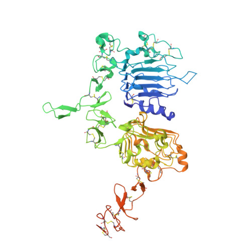
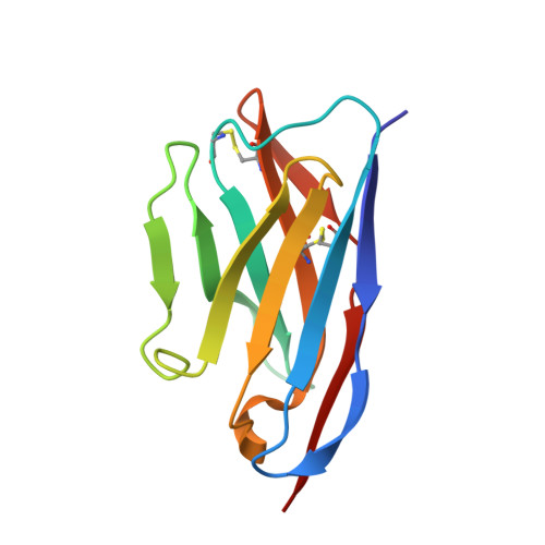
    Purpose: Camelid single-domain antibody-fragments (sdAb) have beneficial pharmacokinetic properties, and those targeted to HER2 can be used for imaging of HER2-overexpressing cancer. Labeled with a therapeutic radionuclide, they may be used for HER2-targeted therapy. Here, we describe the generation of a 131 I-labeled sdAb as a theranostic drug to treat HER2-overexpressing cancer. Experimental Design: Anti-HER2 sdAb 2Rs15d was labeled with 131 I using [ 131 I]SGMIB and evaluated in vitro Biodistribution was evaluated in two HER2 + murine xenograft models by micro-SPECT/CT imaging and at necropsy, and under challenge with trastuzumab and pertuzumab. The therapeutic potential of [ 131 I]SGMIB-2Rs15d was investigated in two HER2 + tumor mouse models. A single-dose toxicity study was performed in mice using unlabeled [ 127 I]SGMIB-sdAb at 1.4 mg/kg. The structure of the 2Rs15d-HER2 complex was determined by X-ray crystallography. Results: [ 131 I]SGMIB-2Rs15d bound specifically to HER2 + cells ( K d = 4.74 ± 0.39 nmol/L). High and specific tumor uptake was observed in both BT474/M1 and SKOV-3 tumor xenografted mice and surpassed kidney levels by 3 hours. Extremely low uptake values were observed in other normal tissues at all time points. The crystal structure revealed that 2Rs15d recognizes HER2 Domain 1, consistent with the lack of competition with trastuzumab and pertuzumab observed in vivo [ 131 I]SGMIB-2Rs15d alone, or in combination with trastuzumab, extended median survival significantly. No toxicity was observed after injecting [ 127 I]SGMIB-2Rs15d. Conclusions: These findings demonstrate the theranostic potential of [ 131 I]SGMIB-2Rs15d. An initial scan using low radioactive [*I]SGMIB-2Rs15d allows patient selection and dosimetry calculations for subsequent therapeutic [ 131 I]SGMIB-2Rs15d and could thereby impact therapy outcome on HER2 + breast cancer patients. Clin Cancer Res; 23(21); 6616-28. ©2017 AACR .


  • Organizational Affiliation

    In Vivo Cellular and Molecular Imaging Laboratory, Vrije Universiteit Brussel, Brussels, Belgium. mdhuyvet@vub.ac.be.


Macromolecules
Find similar proteins by:  (by identity cutoff)  |  3D Structure
Entity ID: 1
MoleculeChains Sequence LengthOrganismDetailsImage
Receptor tyrosine-protein kinase erbB-2622Homo sapiensMutation(s): 0 
Gene Names: ERBB2HER2MLN19NEUNGL
EC: 2.7.10.1
UniProt & NIH Common Fund Data Resources
Find proteins for P04626 (Homo sapiens)
Explore P04626 
Go to UniProtKB:  P04626
PHAROS:  P04626
GTEx:  ENSG00000141736 
Entity Groups  
Sequence Clusters30% Identity50% Identity70% Identity90% Identity95% Identity100% Identity
UniProt GroupP04626
Sequence Annotations
Expand
  • Reference Sequence
Find similar proteins by:  (by identity cutoff)  |  3D Structure
Entity ID: 2
MoleculeChains Sequence LengthOrganismDetailsImage
Nanobody 2Rs15d115Camelus dromedariusMutation(s): 0 
Entity Groups  
Sequence Clusters30% Identity50% Identity70% Identity90% Identity95% Identity100% Identity
Sequence Annotations
Expand
  • Reference Sequence
Oligosaccharides

Help

Entity ID: 3
MoleculeChains Length2D Diagram Glycosylation3D Interactions
2-acetamido-2-deoxy-beta-D-glucopyranose-(1-4)-2-acetamido-2-deoxy-beta-D-glucopyranose
C
2N-Glycosylation
Glycosylation Resources
GlyTouCan:  G42666HT
GlyCosmos:  G42666HT
GlyGen:  G42666HT
Experimental Data & Validation

Experimental Data

  • Method: X-RAY DIFFRACTION
  • Resolution: 2.25 Å
  • R-Value Free: 0.220 
  • R-Value Work: 0.185 
  • R-Value Observed: 0.187 
  • Space Group: P 21 21 21
Unit Cell:
Length ( Å )Angle ( ˚ )
a = 66.52α = 90
b = 113.52β = 90
c = 137.43γ = 90
Software Package:
Software NamePurpose
PHENIXrefinement
PDB_EXTRACTdata extraction
XDSdata scaling
XDSdata reduction
PHASERphasing

Structure Validation

View Full Validation Report


Ligand Structure Quality Assessment 


Entry History & Funding Information

Deposition Data


Funding OrganizationLocationGrant Number
FWO-VlaanderenBelgium--
Innoviris.BrusselsBelgium--
Stichting Tegen KankerBelgium--
BAEFBelgium--
Germaine Eisendrath-Dubois FoundationBelgium--

Revision History  (Full details and data files)

  • Version 1.0: 2017-08-09
    Type: Initial release
  • Version 1.1: 2017-10-11
    Changes: Database references
  • Version 1.2: 2017-11-15
    Changes: Database references
  • Version 2.0: 2020-07-29
    Type: Remediation
    Reason: Carbohydrate remediation
    Changes: Advisory, Atomic model, Data collection, Derived calculations, Structure summary
  • Version 2.1: 2024-01-17
    Changes: Data collection, Database references, Refinement description, Structure summary