5LMF

Structure of C-terminal domain from S. cerevisiae Pat1 decapping activator bound to Dcp2 HLM3 peptide (region 484-500)


Experimental Data Snapshot

  • Method: X-RAY DIFFRACTION
  • Resolution: 2.15 Å
  • R-Value Free: 0.246 
  • R-Value Work: 0.212 
  • R-Value Observed: 0.213 

wwPDB Validation   3D Report Full Report


This is version 1.5 of the entry. See complete history


Literature

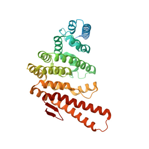
A unique surface on Pat1 C-terminal domain directly interacts with Dcp2 decapping enzyme and Xrn1 5'-3' mRNA exonuclease in yeast.

Charenton, C.Gaudon-Plesse, C.Fourati, Z.Taverniti, V.Back, R.Kolesnikova, O.Seraphin, B.Graille, M.

(2017) Proc Natl Acad Sci U S A 114: E9493-E9501

  • DOI: https://doi.org/10.1073/pnas.1711680114
  • Primary Citation of Related Structures:  
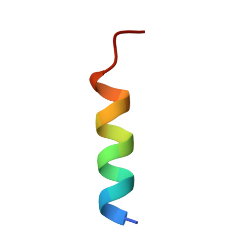
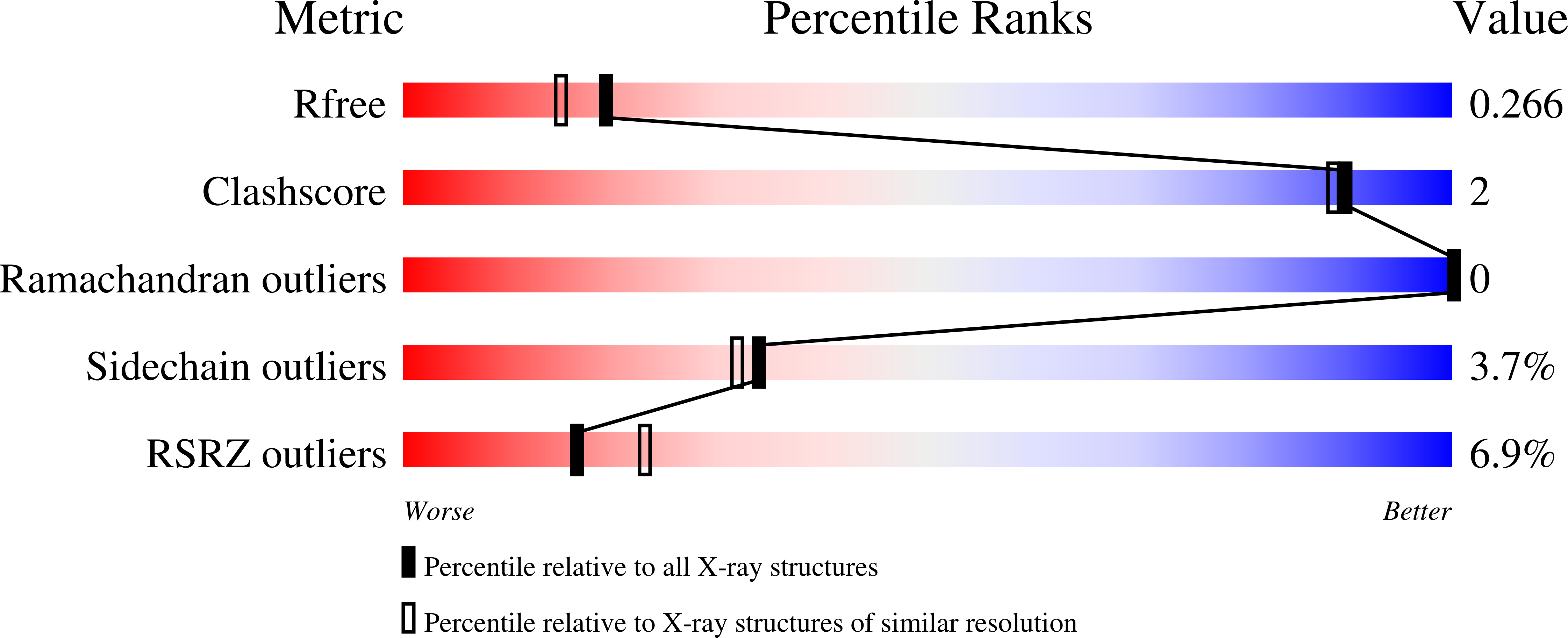
    5LM5, 5LMF, 5LMG

  • PubMed Abstract: 

    The Pat1 protein is a central player of eukaryotic mRNA decay that has also been implicated in translational control. It is commonly considered a central platform responsible for the recruitment of several RNA decay factors. We demonstrate here that a yeast-specific C-terminal region from Pat1 interacts with several short motifs, named helical leucine-rich motifs (HLMs), spread in the long C-terminal region of yeast Dcp2 decapping enzyme. Structures of Pat1-HLM complexes reveal the basis for HLM recognition by Pat1. We also identify a HLM present in yeast Xrn1, the main 5'-3' exonuclease involved in mRNA decay. We show further that the ability of yeast Pat1 to bind HLMs is required for efficient growth and normal mRNA decay. Overall, our analyses indicate that yeast Pat1 uses a single binding surface to successively recruit several mRNA decay factors and show that interaction between those factors is highly polymorphic between species.


  • Organizational Affiliation

    Laboratoire de Biochimie, Ecole Polytechnique, CNRS, Université Paris-Saclay, 91128 Palaiseau cedex, France.


Macromolecules
Find similar proteins by:  (by identity cutoff)  |  3D Structure
Entity ID: 1
MoleculeChains Sequence LengthOrganismDetailsImage
DNA topoisomerase 2-associated protein PAT1
A, B
369Saccharomyces cerevisiaeMutation(s): 0 
Gene Names: PAT1MRT1YCR077CYCR77C
UniProt
Find proteins for P25644 (Saccharomyces cerevisiae (strain ATCC 204508 / S288c))
Explore P25644 
Go to UniProtKB:  P25644
Entity Groups  
Sequence Clusters30% Identity50% Identity70% Identity90% Identity95% Identity100% Identity
UniProt GroupP25644
Sequence Annotations
Expand
  • Reference Sequence

Find similar proteins by:  Sequence   |   3D Structure  

Entity ID: 2
MoleculeChains Sequence LengthOrganismDetailsImage
mRNA decapping protein 2
C, D
17Saccharomyces cerevisiaeMutation(s): 0 
UniProt
Find proteins for P53550 (Saccharomyces cerevisiae (strain ATCC 204508 / S288c))
Explore P53550 
Go to UniProtKB:  P53550
Entity Groups  
Sequence Clusters30% Identity50% Identity70% Identity90% Identity95% Identity100% Identity
UniProt GroupP53550
Sequence Annotations
Expand
  • Reference Sequence
Small Molecules
Ligands 3 Unique
IDChains Name / Formula / InChI Key2D Diagram3D Interactions
EDO
Query on EDO

Download Ideal Coordinates CCD File 
G [auth A]
H [auth A]
I [auth A]
K [auth B]
L [auth B]
G [auth A],
H [auth A],
I [auth A],
K [auth B],
L [auth B],
M [auth B],
N [auth B],
O [auth B],
P [auth B]
1,2-ETHANEDIOL
C2 H6 O2
LYCAIKOWRPUZTN-UHFFFAOYSA-N
CL
Query on CL

Download Ideal Coordinates CCD File 
F [auth A],
J [auth B]
CHLORIDE ION
Cl
VEXZGXHMUGYJMC-UHFFFAOYSA-M
MG
Query on MG

Download Ideal Coordinates CCD File 
E [auth A]MAGNESIUM ION
Mg
JLVVSXFLKOJNIY-UHFFFAOYSA-N
Experimental Data & Validation

Experimental Data

  • Method: X-RAY DIFFRACTION
  • Resolution: 2.15 Å
  • R-Value Free: 0.246 
  • R-Value Work: 0.212 
  • R-Value Observed: 0.213 
  • Space Group: I 2 2 2
Unit Cell:
Length ( Å )Angle ( ˚ )
a = 98.36α = 90
b = 122.92β = 90
c = 126.97γ = 90
Software Package:
Software NamePurpose
BUSTER-TNTrefinement
PDB_EXTRACTdata extraction
XSCALEdata scaling
MOLREPphasing
XDSdata reduction

Structure Validation

View Full Validation Report


Entry History 

Deposition Data

Revision History  (Full details and data files)

  • Version 1.0: 2017-08-16
    Type: Initial release
  • Version 1.1: 2017-11-01
    Changes: Database references
  • Version 1.2: 2017-11-08
    Changes: Database references
  • Version 1.3: 2017-11-15
    Changes: Database references
  • Version 1.4: 2018-09-26
    Changes: Data collection, Derived calculations, Structure summary
  • Version 1.5: 2024-01-10
    Changes: Data collection, Database references, Derived calculations, Refinement description