5KQS

Structure of NS5 methyltransferase from Zika virus bound to S-adenosylmethionine and 7-methyl-guanosine-5'-diphosphate


Experimental Data Snapshot

  • Method: X-RAY DIFFRACTION
  • Resolution: 1.50 Å
  • R-Value Free: 0.179 
  • R-Value Work: 0.159 
  • R-Value Observed: 0.160 

wwPDB Validation   3D Report Full Report


Ligand Structure Quality Assessment 


This is version 1.3 of the entry. See complete history


Literature

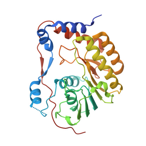
Structures of NS5 Methyltransferase from Zika Virus.

Coloma, J.Jain, R.Rajashankar, K.R.Garcia-Sastre, A.Aggarwal, A.K.

(2016) Cell Rep 16: 3097-3102

  • DOI: https://doi.org/10.1016/j.celrep.2016.08.091
  • Primary Citation of Related Structures:  
    5KQR, 5KQS

  • PubMed Abstract: 

    The Zika virus (ZIKV) poses a major public health emergency. To aid in the development of antivirals, we present two high-resolution crystal structures of the ZIKV NS5 methyltransferase: one bound to S-adenosylmethionine (SAM) and the other bound to SAM and 7-methyl guanosine diphosphate (7-MeGpp). We identify features of ZIKV NS5 methyltransferase that lend to structure-based antiviral drug discovery. Specifically, SAM analogs with functionalities on the Cβ atom of the methionine portion of the molecules that occupy the RNA binding tunnel may provide better specificity relative to human RNA methyltransferases.


  • Organizational Affiliation

    Department of Pharmacological Sciences, Icahn School of Medicine at Mount Sinai, New York, NY 10029, USA.


Macromolecules
Find similar proteins by:  (by identity cutoff)  |  3D Structure
Entity ID: 1
MoleculeChains Sequence LengthOrganismDetailsImage
Methyltransferase268Zika virusMutation(s): 0 
UniProt
Find proteins for A0A024B7W1 (Zika virus (isolate ZIKV/Human/French Polynesia/10087PF/2013))
Explore A0A024B7W1 
Go to UniProtKB:  A0A024B7W1
Entity Groups  
Sequence Clusters30% Identity50% Identity70% Identity90% Identity95% Identity100% Identity
UniProt GroupA0A024B7W1
Sequence Annotations
Expand
  • Reference Sequence
Small Molecules
Ligands 5 Unique
IDChains Name / Formula / InChI Key2D Diagram3D Interactions
M7G
Query on M7G

Download Ideal Coordinates CCD File 
B [auth A]7N-METHYL-8-HYDROGUANOSINE-5'-DIPHOSPHATE
C11 H18 N5 O11 P2
SBASPRRECYVBRF-KQYNXXCUSA-O
SAM
Query on SAM

Download Ideal Coordinates CCD File 
H [auth A]S-ADENOSYLMETHIONINE
C15 H22 N6 O5 S
MEFKEPWMEQBLKI-FCKMPRQPSA-N
PO4
Query on PO4

Download Ideal Coordinates CCD File 
I [auth A]PHOSPHATE ION
O4 P
NBIIXXVUZAFLBC-UHFFFAOYSA-K
GOL
Query on GOL

Download Ideal Coordinates CCD File 
C [auth A],
D [auth A],
E [auth A],
F [auth A]
GLYCEROL
C3 H8 O3
PEDCQBHIVMGVHV-UHFFFAOYSA-N
ACT
Query on ACT

Download Ideal Coordinates CCD File 
G [auth A]ACETATE ION
C2 H3 O2
QTBSBXVTEAMEQO-UHFFFAOYSA-M
Experimental Data & Validation

Experimental Data

  • Method: X-RAY DIFFRACTION
  • Resolution: 1.50 Å
  • R-Value Free: 0.179 
  • R-Value Work: 0.159 
  • R-Value Observed: 0.160 
  • Space Group: C 1 2 1
Unit Cell:
Length ( Å )Angle ( ˚ )
a = 129.34α = 90
b = 77.48β = 104.4
c = 37.03γ = 90
Software Package:
Software NamePurpose
PHENIXrefinement
MOSFLMdata reduction
Aimlessdata scaling
PDB_EXTRACTdata extraction
Auto-Rickshawphasing

Structure Validation

View Full Validation Report


Ligand Structure Quality Assessment 


Entry History 

Deposition Data

Revision History  (Full details and data files)

  • Version 1.0: 2016-09-14
    Type: Initial release
  • Version 1.1: 2017-01-04
    Changes: Database references
  • Version 1.2: 2022-08-03
    Changes: Database references, Structure summary
  • Version 1.3: 2023-10-04
    Changes: Data collection, Refinement description