5JS7

Structural model of a apo G-protein alpha subunit determined with NMR residual dipolar couplings and SAXS


Experimental Data Snapshot

  • Method: SOLUTION NMR
  • Conformers Calculated: 50 
  • Conformers Submitted: 10 
  • Selection Criteria: structures with the least restraint violations 

wwPDB Validation   3D Report Full Report


This is version 1.4 of the entry. See complete history


Literature

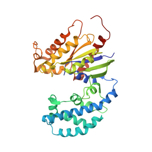
Conformational dynamics of a G-protein alpha subunit is tightly regulated by nucleotide binding.

Goricanec, D.Stehle, R.Egloff, P.Grigoriu, S.Pluckthun, A.Wagner, G.Hagn, F.

(2016) Proc Natl Acad Sci U S A 113: E3629-E3638

  • DOI: https://doi.org/10.1073/pnas.1604125113
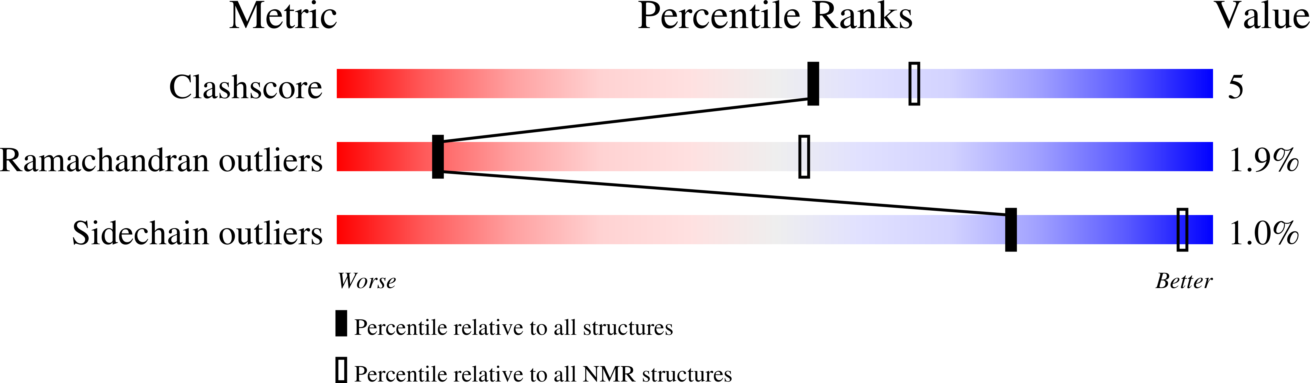
  • Primary Citation of Related Structures:  
    5JS7, 5JS8

  • PubMed Abstract: 

    Heterotrimeric G proteins play a pivotal role in the signal-transduction pathways initiated by G-protein-coupled receptor (GPCR) activation. Agonist-receptor binding causes GDP-to-GTP exchange and dissociation of the Gα subunit from the heterotrimeric G protein, leading to downstream signaling. Here, we studied the internal mobility of a G-protein α subunit in its apo and nucleotide-bound forms and characterized their dynamical features at multiple time scales using solution NMR, small-angle X-ray scattering, and molecular dynamics simulations. We find that binding of GTP analogs leads to a rigid and closed arrangement of the Gα subdomain, whereas the apo and GDP-bound forms are considerably more open and dynamic. Furthermore, we were able to detect two conformational states of the Gα Ras domain in slow exchange whose populations are regulated by binding to nucleotides and a GPCR. One of these conformational states, the open state, binds to the GPCR; the second conformation, the closed state, shows no interaction with the receptor. Binding to the GPCR stabilizes the open state. This study provides an in-depth analysis of the conformational landscape and the switching function of a G-protein α subunit and the influence of a GPCR in that landscape.


  • Organizational Affiliation

    Institute for Advanced Study at the Department of Chemistry, Technische Universität München, 85747 Garching, Germany; Center for Integrated Protein Science Munich at the Department of Chemistry, Technische Universität München, 85747 Garching, Germany; Institute of Structural Biology, Helmholtz Zentrum München, 85764 Neuherberg, Germany;


Macromolecules
Find similar proteins by:  (by identity cutoff)  |  3D Structure
Entity ID: 1
MoleculeChains Sequence LengthOrganismDetailsImage
Guanine nucleotide-binding protein G(i) subunit alpha-1326Homo sapiensMutation(s): 0 
Gene Names: GNAI1
UniProt & NIH Common Fund Data Resources
Find proteins for P63096 (Homo sapiens)
Explore P63096 
Go to UniProtKB:  P63096
PHAROS:  P63096
GTEx:  ENSG00000127955 
Entity Groups  
Sequence Clusters30% Identity50% Identity70% Identity90% Identity95% Identity100% Identity
UniProt GroupP63096
Sequence Annotations
Expand
  • Reference Sequence
Experimental Data & Validation

Experimental Data

  • Method: SOLUTION NMR
  • Conformers Calculated: 50 
  • Conformers Submitted: 10 
  • Selection Criteria: structures with the least restraint violations 

Structure Validation

View Full Validation Report


Entry History & Funding Information

Deposition Data


Funding OrganizationLocationGrant Number
National Institutes of HealthUnited StatesGM075879
National Institutes of HealthUnited StatesGM047467
National Institutes of HealthUnited StatesGM094608
National Institutes of HealthUnited StatesEB002026
European CommissionGermany291763
Center for Integrated Protein Science MunichGermany--
HFSPFranceLT-000297/2011

Revision History  (Full details and data files)

  • Version 1.0: 2016-06-29
    Type: Initial release
  • Version 1.1: 2016-07-06
    Changes: Database references
  • Version 1.2: 2018-01-31
    Changes: Author supporting evidence
  • Version 1.3: 2019-05-08
    Changes: Data collection
  • Version 1.4: 2023-06-14
    Changes: Data collection, Database references, Other