5HZF

Single Chain Recombinant Globular Head of the Complement System Protein C1q in complex with magnesium


Experimental Data Snapshot

  • Method: X-RAY DIFFRACTION
  • Resolution: 1.55 Å
  • R-Value Free: 0.186 
  • R-Value Work: 0.169 
  • R-Value Observed: 0.170 

wwPDB Validation   3D Report Full Report


This is version 2.2 of the entry. See complete history


Literature

Structural and Functional Characterization of a Single-Chain Form of the Recognition Domain of Complement Protein C1q.

Moreau, C.Bally, I.Chouquet, A.Bottazzi, B.Ghebrehiwet, B.Gaboriaud, C.Thielens, N.

(2016) Front Immunol 7: 79-79

  • DOI: https://doi.org/10.3389/fimmu.2016.00079
  • Primary Citation of Related Structures:  
    5HZF

  • PubMed Abstract: 

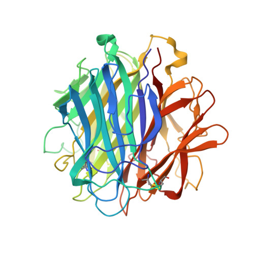
    Complement C1q is a soluble pattern recognition molecule comprising six heterotrimeric subunits assembled from three polypeptide chains (A-C). Each heterotrimer forms a collagen-like stem prolonged by a globular recognition domain. These recognition domains sense a wide variety of ligands, including pathogens and altered-self components. Ligand recognition is either direct or mediated by immunoglobulins or pentraxins. Multivalent binding of C1q to its targets triggers immune effector mechanisms mediated via its collagen-like stems. The induced immune response includes activation of the classical complement pathway and enhancement of the phagocytosis of the recognized target. We report here, the first production of a single-chain recombinant form of human C1q globular region (C1q-scGR). The three monomers have been linked in tandem to generate a single continuous polypeptide, based on a strategy previously used for adiponectin, a protein structurally related to C1q. The resulting C1q-scGR protein was produced at high yield in stably transfected 293-F mammalian cells. Recombinant C1q-scGR was correctly folded, as demonstrated by its X-ray crystal structure solved at a resolution of 1.35 Å. Its interaction properties were assessed by surface plasmon resonance analysis using the following physiological C1q ligands: the receptor for C1q globular heads, the long pentraxin PTX3, calreticulin, and heparin. The 3D structure and the binding properties of C1q-scGR were similar to those of the three-chain fragment generated by collagenase digestion of serum-derived C1q. Comparison of the interaction properties of the fragments with those of native C1q provided insights into the avidity component associated with the hexameric assembly of C1q. The interest of this functional recombinant form of the recognition domains of C1q in basic research and its potential biomedical applications are discussed.


  • Organizational Affiliation

    IBS, University of Grenoble Alpes, Grenoble, France; CNRS, IBS, Grenoble, France; IBS, CEA, Grenoble, France.


Macromolecules
Find similar proteins by:  (by identity cutoff)  |  3D Structure
Entity ID: 1
MoleculeChains Sequence LengthOrganismDetailsImage
Complement C1q subcomponent subunit A,Complement C1q subcomponent subunit C,Complement C1q subcomponent subunit B410Homo sapiensMutation(s): 0 
Gene Names: C1QAC1QCC1QGC1QB
UniProt & NIH Common Fund Data Resources
Find proteins for P02747 (Homo sapiens)
Explore P02747 
Go to UniProtKB:  P02747
PHAROS:  P02747
GTEx:  ENSG00000159189 
Find proteins for P02746 (Homo sapiens)
Explore P02746 
Go to UniProtKB:  P02746
PHAROS:  P02746
Find proteins for P02745 (Homo sapiens)
Explore P02745 
Go to UniProtKB:  P02745
PHAROS:  P02745
GTEx:  ENSG00000173372 
Entity Groups  
Sequence Clusters30% Identity50% Identity70% Identity90% Identity95% Identity100% Identity
UniProt GroupsP02747P02745P02746
Sequence Annotations
Expand
  • Reference Sequence
Small Molecules
Ligands 1 Unique
IDChains Name / Formula / InChI Key2D Diagram3D Interactions
MG
Query on MG

Download Ideal Coordinates CCD File 
B [auth A]MAGNESIUM ION
Mg
JLVVSXFLKOJNIY-UHFFFAOYSA-N
Experimental Data & Validation

Experimental Data

  • Method: X-RAY DIFFRACTION
  • Resolution: 1.55 Å
  • R-Value Free: 0.186 
  • R-Value Work: 0.169 
  • R-Value Observed: 0.170 
  • Space Group: C 1 2 1
Unit Cell:
Length ( Å )Angle ( ˚ )
a = 80.996α = 90
b = 52.911β = 115.19
c = 89.894γ = 90
Software Package:
Software NamePurpose
PHENIXrefinement
PHASERphasing
XDSdata reduction
XSCALEdata scaling

Structure Validation

View Full Validation Report


Entry History & Funding Information

Deposition Data


Funding OrganizationLocationGrant Number
French National Research AgencyFranceANR 09-PIRI-0021

Revision History  (Full details and data files)

  • Version 1.0: 2016-03-02
    Type: Initial release
  • Version 1.1: 2016-03-30
    Changes: Database references
  • Version 2.0: 2017-08-30
    Changes: Atomic model, Author supporting evidence
  • Version 2.1: 2019-06-12
    Changes: Data collection, Structure summary
  • Version 2.2: 2024-01-10
    Changes: Data collection, Database references, Refinement description