5HCD

Ternary complex of human Complement C5 with Ornithodoros moubata OmCI and Rhipicephalus microplus RaCI2


Experimental Data Snapshot

  • Method: X-RAY DIFFRACTION
  • Resolution: 2.98 Å
  • R-Value Free: 0.282 
  • R-Value Work: 0.257 
  • R-Value Observed: 0.259 

wwPDB Validation   3D Report Full Report


This is version 2.1 of the entry. See complete history


Literature

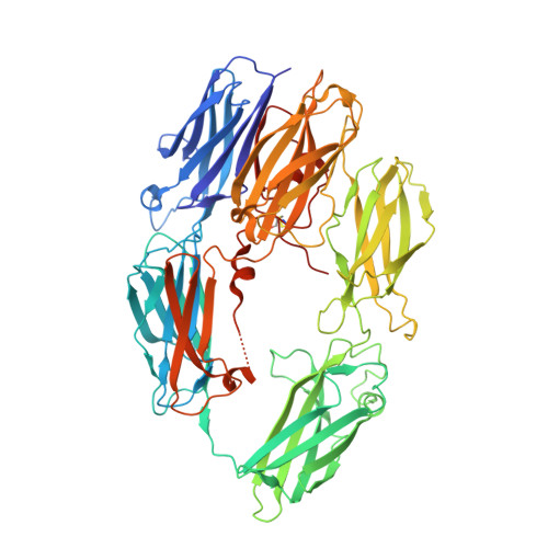
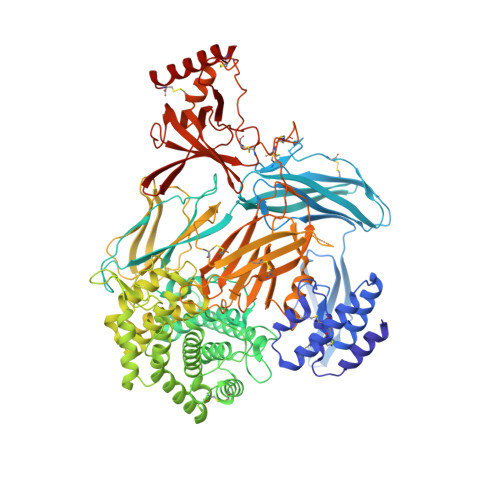
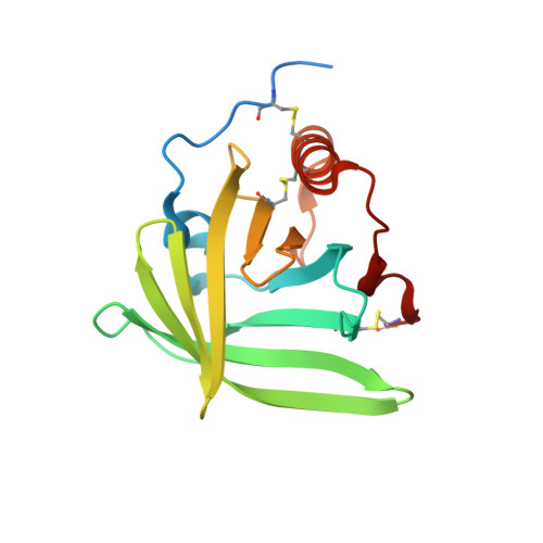
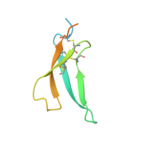
Structural basis for therapeutic inhibition of complement C5.

Jore, M.M.Johnson, S.Sheppard, D.Barber, N.M.Li, Y.I.Nunn, M.A.Elmlund, H.Lea, S.M.

(2016) Nat Struct Mol Biol 23: 378-386

  • DOI: https://doi.org/10.1038/nsmb.3196
  • Primary Citation of Related Structures:  
    5HCC, 5HCD, 5IEC

  • PubMed Abstract: 

    Activation of complement C5 generates the potent anaphylatoxin C5a and leads to pathogen lysis, inflammation and cell damage. The therapeutic potential of C5 inhibition has been demonstrated by eculizumab, one of the world's most expensive drugs. However, the mechanism of C5 activation by C5 convertases remains elusive, thus limiting development of therapeutics. Here we identify and characterize a new protein family of tick-derived C5 inhibitors. Structures of C5 in complex with the new inhibitors, the phase I and phase II inhibitor OmCI, or an eculizumab Fab reveal three distinct binding sites on C5 that all prevent activation of C5. The positions of the inhibitor-binding sites and the ability of all three C5-inhibitor complexes to competitively inhibit the C5 convertase conflict with earlier steric-inhibition models, thus suggesting that a priming event is needed for activation.


  • Organizational Affiliation

    Sir William Dunn School of Pathology, University of Oxford, Oxford, UK.


Macromolecules
Find similar proteins by:  (by identity cutoff)  |  3D Structure
Entity ID: 1
MoleculeChains Sequence LengthOrganismDetailsImage
Complement C5A [auth B]656Homo sapiensMutation(s): 0 
UniProt & NIH Common Fund Data Resources
Find proteins for P01031 (Homo sapiens)
Explore P01031 
Go to UniProtKB:  P01031
PHAROS:  P01031
GTEx:  ENSG00000106804 
Entity Groups  
Sequence Clusters30% Identity50% Identity70% Identity90% Identity95% Identity100% Identity
UniProt GroupP01031
Sequence Annotations
Expand
  • Reference Sequence
Find similar proteins by:  (by identity cutoff)  |  3D Structure
Entity ID: 2
MoleculeChains Sequence LengthOrganismDetailsImage
Complement C5B [auth A]998Homo sapiensMutation(s): 0 
UniProt & NIH Common Fund Data Resources
Find proteins for P01031 (Homo sapiens)
Explore P01031 
Go to UniProtKB:  P01031
PHAROS:  P01031
GTEx:  ENSG00000106804 
Entity Groups  
Sequence Clusters30% Identity50% Identity70% Identity90% Identity95% Identity100% Identity
UniProt GroupP01031
Sequence Annotations
Expand
  • Reference Sequence
Find similar proteins by:  (by identity cutoff)  |  3D Structure
Entity ID: 3
MoleculeChains Sequence LengthOrganismDetailsImage
Complement inhibitor165Ornithodoros moubataMutation(s): 2 
Gene Names: CI
UniProt
Find proteins for Q5YD59 (Ornithodoros moubata)
Explore Q5YD59 
Go to UniProtKB:  Q5YD59
Entity Groups  
Sequence Clusters30% Identity50% Identity70% Identity90% Identity95% Identity100% Identity
UniProt GroupQ5YD59
Sequence Annotations
Expand
  • Reference Sequence
Find similar proteins by:  (by identity cutoff)  |  3D Structure
Entity ID: 4
MoleculeChains Sequence LengthOrganismDetailsImage
Rhipicephalus microplus RaCI280Rhipicephalus microplusMutation(s): 0 
UniProt
Find proteins for A0A158RFT4 (Rhipicephalus microplus)
Explore A0A158RFT4 
Go to UniProtKB:  A0A158RFT4
Entity Groups  
Sequence Clusters30% Identity50% Identity70% Identity90% Identity95% Identity100% Identity
UniProt GroupA0A158RFT4
Sequence Annotations
Expand
  • Reference Sequence
Oligosaccharides

Help

Entity ID: 5
MoleculeChains Length2D Diagram Glycosylation3D Interactions
2-acetamido-2-deoxy-beta-D-glucopyranose-(1-4)-2-acetamido-2-deoxy-beta-D-glucopyranose
E
2N-Glycosylation
Glycosylation Resources
GlyTouCan:  G42666HT
GlyCosmos:  G42666HT
GlyGen:  G42666HT
Small Molecules
Ligands 1 Unique
IDChains Name / Formula / InChI Key2D Diagram3D Interactions
CYS
Query on CYS

Download Ideal Coordinates CCD File 
F [auth A]CYSTEINE
C3 H7 N O2 S
XUJNEKJLAYXESH-REOHCLBHSA-N
Experimental Data & Validation

Experimental Data

  • Method: X-RAY DIFFRACTION
  • Resolution: 2.98 Å
  • R-Value Free: 0.282 
  • R-Value Work: 0.257 
  • R-Value Observed: 0.259 
  • Space Group: P 21 21 21
Unit Cell:
Length ( Å )Angle ( ˚ )
a = 105.954α = 90
b = 140.207β = 90
c = 209.792γ = 90
Software Package:
Software NamePurpose
PHENIXrefinement
xia2data reduction
Aimlessdata scaling
PHASERphasing

Structure Validation

View Full Validation Report


Entry History & Funding Information

Deposition Data


Funding OrganizationLocationGrant Number
Wellcome TrustUnited Kingdom100298
Netherlands Organisation for Scientific ResearchNetherlands825.11.030

Revision History  (Full details and data files)

  • Version 1.0: 2016-03-30
    Type: Initial release
  • Version 1.1: 2016-04-06
    Changes: Database references
  • Version 1.2: 2016-05-11
    Changes: Database references
  • Version 2.0: 2020-07-29
    Type: Remediation
    Reason: Carbohydrate remediation
    Changes: Advisory, Atomic model, Data collection, Derived calculations, Structure summary
  • Version 2.1: 2024-01-10
    Changes: Data collection, Database references, Refinement description, Structure summary