5FT8

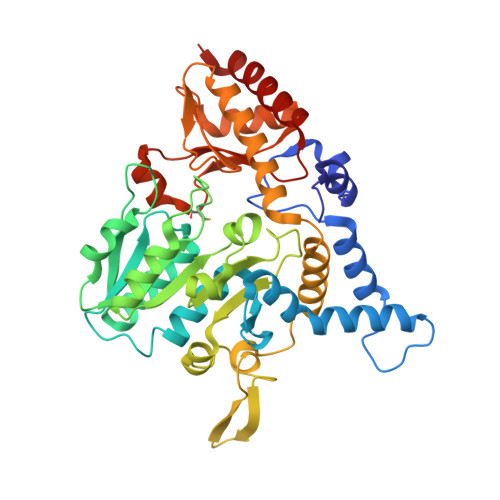
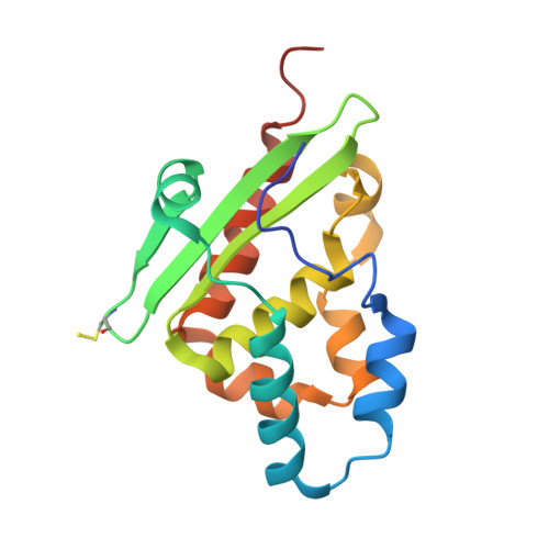
Crystal structure of the complex between the cysteine desulfurase CsdA and the sulfur-acceptor CsdE in the persulfurated state at 2.50 Angstroem resolution


Experimental Data Snapshot

  • Method: X-RAY DIFFRACTION
  • Resolution: 2.50 Å
  • R-Value Free: 0.279 
  • R-Value Work: 0.254 
  • R-Value Observed: 0.254 

wwPDB Validation   3D Report Full Report


Ligand Structure Quality Assessment 


This is version 2.1 of the entry. See complete history


Literature

Mechanism of Sulfur Transfer Across Protein-Protein Interfaces: The Cysteine Desulfurase Model System

Fernandez, F.J.Arda, A.Lopez-Estepa, M.Aranda, J.Penya-Soler, E.Garces, F.Quintana, J.F.Round, A.Campos-Oliva, R.Bruix, M.Coll, M.Tunon, I.Jimenez-Barbero, J.Vega, M.C.

(2016) ACS Catal 6: 3975


Macromolecules
Find similar proteins by:  (by identity cutoff)  |  3D Structure
Entity ID: 1
MoleculeChains Sequence LengthOrganismDetailsImage
Cysteine desulfurase CsdA
A, C, E, G, I
A, C, E, G, I, K, M, O
403Escherichia coli K-12Mutation(s): 0 
Gene Names: csdAygdJb2810JW2781
EC: 2.8.1.7 (PDB Primary Data), 3.13.1 (PDB Primary Data), 4.4.1.16 (PDB Primary Data)
UniProt
Find proteins for Q46925 (Escherichia coli (strain K12))
Explore Q46925 
Go to UniProtKB:  Q46925
Entity Groups  
Sequence Clusters30% Identity50% Identity70% Identity90% Identity95% Identity100% Identity
UniProt GroupQ46925
Sequence Annotations
Expand
  • Reference Sequence
Find similar proteins by:  (by identity cutoff)  |  3D Structure
Entity ID: 2
MoleculeChains Sequence LengthOrganismDetailsImage
Sulfur acceptor protein CsdE154Escherichia coli K-12Mutation(s): 0 
Gene Names: csdEygdKb2811JW2782
UniProt
Find proteins for P0AGF2 (Escherichia coli (strain K12))
Explore P0AGF2 
Go to UniProtKB:  P0AGF2
Entity Groups  
Sequence Clusters30% Identity50% Identity70% Identity90% Identity95% Identity100% Identity
UniProt GroupP0AGF2
Sequence Annotations
Expand
  • Reference Sequence
Small Molecules
Ligands 3 Unique
IDChains Name / Formula / InChI Key2D Diagram3D Interactions
PLP
Query on PLP

Download Ideal Coordinates CCD File 
AB [auth E]
BC [auth I]
IC [auth K]
KA [auth C]
KC [auth M]
AB [auth E],
BC [auth I],
IC [auth K],
KA [auth C],
KC [auth M],
MC [auth O],
Q [auth A],
QB [auth G]
PYRIDOXAL-5'-PHOSPHATE
C8 H10 N O6 P
NGVDGCNFYWLIFO-UHFFFAOYSA-N
PEG
Query on PEG

Download Ideal Coordinates CCD File 
BB [auth E]
CB [auth E]
CC [auth I]
DB [auth E]
EB [auth E]
BB [auth E],
CB [auth E],
CC [auth I],
DB [auth E],
EB [auth E],
GA [auth Q],
JC [auth K],
LA [auth C],
MA [auth C],
NA [auth C],
OA [auth C],
R [auth A],
RB [auth G],
S [auth A],
T [auth A],
ZB [auth W]
DI(HYDROXYETHYL)ETHER
C4 H10 O3
MTHSVFCYNBDYFN-UHFFFAOYSA-N
GOL
Query on GOL

Download Ideal Coordinates CCD File 
AA [auth A]
AC [auth W]
BA [auth A]
CA [auth A]
DA [auth A]
AA [auth A],
AC [auth W],
BA [auth A],
CA [auth A],
DA [auth A],
DC [auth I],
EA [auth A],
EC [auth I],
FA [auth A],
FB [auth E],
FC [auth I],
GB [auth E],
GC [auth I],
HA [auth Q],
HB [auth E],
HC [auth I],
IA [auth Q],
IB [auth E],
JA [auth Q],
JB [auth E],
KB [auth E],
LB [auth E],
LC [auth M],
MB [auth E],
NB [auth E],
OB [auth E],
PA [auth C],
PB [auth E],
QA [auth C],
RA [auth C],
SA [auth C],
SB [auth G],
TA [auth C],
TB [auth G],
U [auth A],
UA [auth C],
UB [auth G],
V [auth A],
VA [auth C],
VB [auth G],
W [auth A],
WA [auth C],
WB [auth G],
X [auth A],
XA [auth C],
XB [auth G],
Y [auth A],
YA [auth C],
YB [auth G],
Z [auth A],
ZA [auth S]
GLYCEROL
C3 H8 O3
PEDCQBHIVMGVHV-UHFFFAOYSA-N
Modified Residues  1 Unique
IDChains TypeFormula2D DiagramParent
CSS
Query on CSS
A, C, E, G, I
A, C, E, G, I, K, M, O
L-PEPTIDE LINKINGC3 H7 N O2 S2CYS
Experimental Data & Validation

Experimental Data

  • Method: X-RAY DIFFRACTION
  • Resolution: 2.50 Å
  • R-Value Free: 0.279 
  • R-Value Work: 0.254 
  • R-Value Observed: 0.254 
  • Space Group: P 21 21 21
Unit Cell:
Length ( Å )Angle ( ˚ )
a = 74.863α = 90
b = 115.135β = 90
c = 604.966γ = 90
Software Package:
Software NamePurpose
REFMACrefinement
XDSdata reduction
Aimlessdata scaling
PHASERphasing

Structure Validation

View Full Validation Report


Ligand Structure Quality Assessment 


Entry History 

Revision History  (Full details and data files)

  • Version 1.0: 2016-11-23
    Type: Initial release
  • Version 1.1: 2019-01-02
    Changes: Data collection, Database references, Structure summary
  • Version 2.0: 2022-12-07
    Changes: Advisory, Atomic model, Data collection, Database references, Derived calculations, Other, Polymer sequence, Source and taxonomy, Structure summary
  • Version 2.1: 2024-01-31
    Changes: Data collection, Refinement description