5F9C

Crystal structure of the G121R mutant of human phosphoglucomutase 1


Experimental Data Snapshot

  • Method: X-RAY DIFFRACTION
  • Resolution: 2.50 Å
  • R-Value Free: 0.243 
  • R-Value Work: 0.192 
  • R-Value Observed: 0.194 

wwPDB Validation   3D Report Full Report


This is version 1.2 of the entry. See complete history


Literature

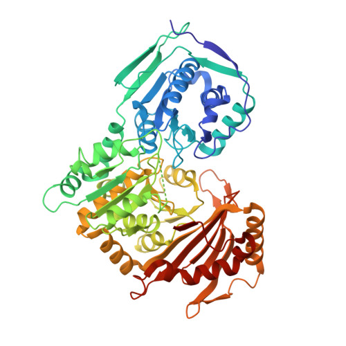
Induced Structural Disorder as a Molecular Mechanism for Enzyme Dysfunction in Phosphoglucomutase 1 Deficiency.

Stiers, K.M.Kain, B.N.Graham, A.C.Beamer, L.J.

(2016) J Mol Biol 428: 1493-1505

  • DOI: https://doi.org/10.1016/j.jmb.2016.02.032
  • Primary Citation of Related Structures:  
    5EPC, 5F9C, 5HSH

  • PubMed Abstract: 

    Human phosphoglucomutase 1 (PGM1) plays a central role in cellular glucose homeostasis, mediating the switch between glycolysis and gluconeogenesis through the conversion of glucose 1-phosphate and glucose 6-phosphate. Recent clinical studies have identified mutations in this enzyme as the cause of PGM1 deficiency, an inborn error of metabolism classified as both a glycogen storage disease and a congenital disorder of glycosylation. Reported here are the first crystal structures of two disease-related missense variants of PGM1, along with the structure of the wild-type enzyme. Two independent glycine-to-arginine substitutions (G121R and G291R), both affecting key active site loops of PGM1, are found to induce regions of structural disorder, as evidenced by a nearly complete loss of electron density for as many as 23 aa. The disordered regions are not contiguous in sequence to the site of mutation, and even cross domain boundaries. Other structural rearrangements include changes in the conformations of loops and side chains, some of which occur nearly 20 Å away from the site of mutation. The induced structural disorder is correlated with increased sensitivity to proteolysis and lower-resolution diffraction, particularly for the G291R variant. Examination of the multi-domain effects of these G➔R mutations establishes a correlation between interdomain interfaces of the enzyme and missense variants of PGM1 associated with disease. These crystal structures provide the first insights into the structural basis of enzyme dysfunction in PGM1 deficiency and highlight a growing role for biophysical characterization of proteins in the field of precision medicine.


  • Organizational Affiliation

    Biochemistry Department, University of Missouri, 117 Schweitzer Hall, Columbia, MO 65211, USA.


Macromolecules
Find similar proteins by:  (by identity cutoff)  |  3D Structure
Entity ID: 1
MoleculeChains Sequence LengthOrganismDetailsImage
Phosphoglucomutase-1
A, B
562Homo sapiensMutation(s): 1 
Gene Names: PGM1
EC: 5.4.2.2
UniProt & NIH Common Fund Data Resources
Find proteins for P36871 (Homo sapiens)
Explore P36871 
Go to UniProtKB:  P36871
PHAROS:  P36871
GTEx:  ENSG00000079739 
Entity Groups  
Sequence Clusters30% Identity50% Identity70% Identity90% Identity95% Identity100% Identity
UniProt GroupP36871
Sequence Annotations
Expand
  • Reference Sequence
Small Molecules
Ligands 3 Unique
IDChains Name / Formula / InChI Key2D Diagram3D Interactions
SO4
Query on SO4

Download Ideal Coordinates CCD File 
D [auth A]
E [auth A]
F [auth A]
I [auth B]
J [auth B]
D [auth A],
E [auth A],
F [auth A],
I [auth B],
J [auth B],
K [auth B],
L [auth B],
M [auth B],
N [auth B]
SULFATE ION
O4 S
QAOWNCQODCNURD-UHFFFAOYSA-L
GOL
Query on GOL

Download Ideal Coordinates CCD File 
G [auth A],
H [auth A]
GLYCEROL
C3 H8 O3
PEDCQBHIVMGVHV-UHFFFAOYSA-N
MG
Query on MG

Download Ideal Coordinates CCD File 
C [auth A],
O [auth B]
MAGNESIUM ION
Mg
JLVVSXFLKOJNIY-UHFFFAOYSA-N
Experimental Data & Validation

Experimental Data

  • Method: X-RAY DIFFRACTION
  • Resolution: 2.50 Å
  • R-Value Free: 0.243 
  • R-Value Work: 0.192 
  • R-Value Observed: 0.194 
  • Space Group: P 41 21 2
Unit Cell:
Length ( Å )Angle ( ˚ )
a = 171.98α = 90
b = 171.98β = 90
c = 99.9γ = 90
Software Package:
Software NamePurpose
PHENIXrefinement
XDSdata reduction
XDSdata scaling

Structure Validation

View Full Validation Report


Entry History 

Deposition Data

  • Released Date: 2016-04-27 
  • Deposition Author(s): Beamer, L.J.

Revision History  (Full details and data files)

  • Version 1.0: 2016-04-27
    Type: Initial release
  • Version 1.1: 2017-11-22
    Changes: Database references, Derived calculations, Refinement description
  • Version 1.2: 2023-09-27
    Changes: Data collection, Database references, Refinement description