5EOC

Crystal structure of Fab C2 in complex with a Cyclic variant of Hepatitis C Virus E2 epitope I


Experimental Data Snapshot

  • Method: X-RAY DIFFRACTION
  • Resolution: 1.98 Å
  • R-Value Free: 0.266 
  • R-Value Work: 0.234 

wwPDB Validation   3D Report Full Report


This is version 1.2 of the entry. See complete history


Literature

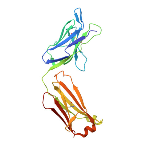
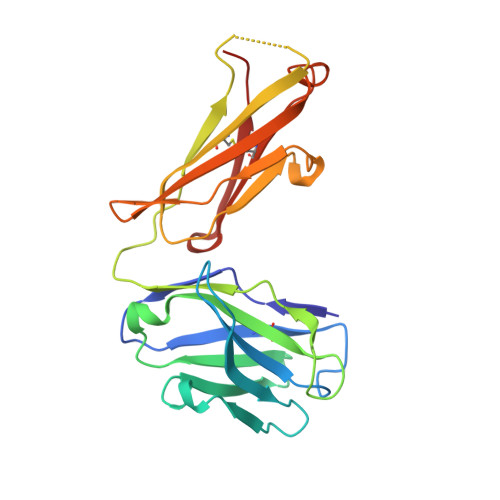
Generation and Characterization of Monoclonal Antibodies against a Cyclic Variant of Hepatitis C Virus E2 Epitope 412-422.

Sandomenico, A.Leonardi, A.Berisio, R.Sanguigno, L.Foca, G.Foca, A.Ruggiero, A.Doti, N.Muscariello, L.Barone, D.Farina, C.Owsianka, A.Vitagliano, L.Patel, A.H.Ruvo, M.

(2016) J Virol 90: 3745-3759

  • DOI: https://doi.org/10.1128/JVI.02397-15
  • Primary Citation of Related Structures:  
    5EOC

  • PubMed Abstract: 

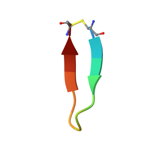
    The hepatitis C virus (HCV) E2 envelope glycoprotein is crucial for virus entry into hepatocytes. A conserved region of E2 encompassing amino acids 412 to 423 (epitope I) and containing Trp420, a residue critical for virus entry, is recognized by several broadly neutralizing antibodies. Peptides embodying this epitope I sequence adopt a β-hairpin conformation when bound to neutralizing monoclonal antibodies (MAbs) AP33 and HCV1. We therefore generated new mouse MAbs that were able to bind to a cyclic peptide containing E2 residues 412 to 422 (C-epitope I) but not to the linear counterpart. These MAbs bound to purified E2 with affinities of about 50 nM, but they were unable to neutralize virus infection. Structural analysis of the complex between C-epitope I and one of our MAbs (C2) showed that the Trp420 side chain is largely buried in the combining site and that the Asn417 side chain, which is glycosylated in E2 and solvent exposed in other complexes, is slightly buried upon C2 binding. Also, the orientation of the cyclic peptide in the antibody-combining site is rotated by 180° compared to the orientations of the other complexes. All these structural features, however, do not explain the lack of neutralization activity. This is instead ascribed to the high degree of selectivity of the new MAbs for the cyclic epitope and to their inability to interact with the epitope in more flexible and extended conformations, which recent data suggest play a role in the mechanisms of neutralization escape.


  • Organizational Affiliation

    Institute of Biostructures and Bioimaging, CNR and CIRPeB, University of Naples Federico II, Naples, Italy.


Macromolecules
Find similar proteins by:  (by identity cutoff)  |  3D Structure
Entity ID: 1
MoleculeChains Sequence LengthOrganismDetailsImage
Fab fragment (Heavy chain)A [auth H]217Mus musculusMutation(s): 0 
Entity Groups  
Sequence Clusters30% Identity50% Identity70% Identity90% Identity95% Identity100% Identity
Sequence Annotations
Expand
  • Reference Sequence
Find similar proteins by:  (by identity cutoff)  |  3D Structure
Entity ID: 2
MoleculeChains Sequence LengthOrganismDetailsImage
Fab fragment (Light chain)B [auth L],
D [auth M]
216Mus musculusMutation(s): 0 
Entity Groups  
Sequence Clusters30% Identity50% Identity70% Identity90% Identity95% Identity100% Identity
Sequence Annotations
Expand
  • Reference Sequence
Find similar proteins by:  (by identity cutoff)  |  3D Structure
Entity ID: 3
MoleculeChains Sequence LengthOrganismDetailsImage
Fab fragment (Heavy chain)C [auth J]217Mus musculusMutation(s): 0 
Entity Groups  
Sequence Clusters30% Identity50% Identity70% Identity90% Identity95% Identity100% Identity
Sequence Annotations
Expand
  • Reference Sequence

Find similar proteins by:  Sequence   |   3D Structure  

Entity ID: 4
MoleculeChains Sequence LengthOrganismDetailsImage
ALA-CYS-GLN-LEU-ILE-ASN-THR-ASN-GLY-SER-TRP-HIS-ILE-CYSE [auth P],
F [auth Q]
14Hepacivirus hominisMutation(s): 0 
UniProt
Find proteins for P27958 (Hepatitis C virus genotype 1a (isolate H77))
Explore P27958 
Go to UniProtKB:  P27958
Entity Groups  
Sequence Clusters30% Identity50% Identity70% Identity90% Identity95% Identity100% Identity
UniProt GroupP27958
Sequence Annotations
Expand
  • Reference Sequence
Experimental Data & Validation

Experimental Data

  • Method: X-RAY DIFFRACTION
  • Resolution: 1.98 Å
  • R-Value Free: 0.266 
  • R-Value Work: 0.234 
  • Space Group: P 1
Unit Cell:
Length ( Å )Angle ( ˚ )
a = 54.972α = 90.35
b = 56.191β = 90.18
c = 77.153γ = 94.98
Software Package:
Software NamePurpose
REFMACrefinement
HKL-2000data reduction
HKL-2000data scaling
PHASERphasing

Structure Validation

View Full Validation Report


Entry History & Funding Information

Deposition Data


Funding OrganizationLocationGrant Number
Ministry of Education, Universities and ResearchItalyRBNE08NKH7_003

Revision History  (Full details and data files)

  • Version 1.0: 2016-02-10
    Type: Initial release
  • Version 1.1: 2016-03-23
    Changes: Database references
  • Version 1.2: 2024-01-10
    Changes: Data collection, Database references, Refinement description