5E3H

Structural Basis for RNA Recognition and Activation of RIG-I


Experimental Data Snapshot

  • Method: X-RAY DIFFRACTION
  • Resolution: 2.70 Å
  • R-Value Free: 0.268 
  • R-Value Work: 0.234 
  • R-Value Observed: 0.236 

wwPDB Validation   3D Report Full Report


Ligand Structure Quality Assessment 


This is version 1.3 of the entry. See complete history


Literature

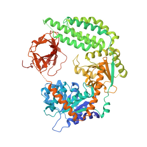
Structural basis of RNA recognition and activation by innate immune receptor RIG-I.

Jiang, F.Ramanathan, A.Miller, M.T.Tang, G.Q.Gale, M.Patel, S.S.Marcotrigiano, J.

(2011) Nature 479: 423-427

  • DOI: https://doi.org/10.1038/nature10537
  • Primary Citation of Related Structures:  
    5E3H

  • PubMed Abstract: 

    Retinoic-acid-inducible gene-I (RIG-I; also known as DDX58) is a cytoplasmic pathogen recognition receptor that recognizes pathogen-associated molecular pattern (PAMP) motifs to differentiate between viral and cellular RNAs. RIG-I is activated by blunt-ended double-stranded (ds)RNA with or without a 5'-triphosphate (ppp), by single-stranded RNA marked by a 5'-ppp and by polyuridine sequences. Upon binding to such PAMP motifs, RIG-I initiates a signalling cascade that induces innate immune defences and inflammatory cytokines to establish an antiviral state. The RIG-I pathway is highly regulated and aberrant signalling leads to apoptosis, altered cell differentiation, inflammation, autoimmune diseases and cancer. The helicase and repressor domains (RD) of RIG-I recognize dsRNA and 5'-ppp RNA to activate the two amino-terminal caspase recruitment domains (CARDs) for signalling. Here, to understand the synergy between the helicase and the RD for RNA binding, and the contribution of ATP hydrolysis to RIG-I activation, we determined the structure of human RIG-I helicase-RD in complex with dsRNA and an ATP analogue. The helicase-RD organizes into a ring around dsRNA, capping one end, while contacting both strands using previously uncharacterized motifs to recognize dsRNA. Small-angle X-ray scattering, limited proteolysis and differential scanning fluorimetry indicate that RIG-I is in an extended and flexible conformation that compacts upon binding RNA. These results provide a detailed view of the role of helicase in dsRNA recognition, the synergy between the RD and the helicase for RNA binding and the organization of full-length RIG-I bound to dsRNA, and provide evidence of a conformational change upon RNA binding. The RIG-I helicase-RD structure is consistent with dsRNA translocation without unwinding and cooperative binding to RNA. The structure yields unprecedented insight into innate immunity and has a broader impact on other areas of biology, including RNA interference and DNA repair, which utilize homologous helicase domains within DICER and FANCM.


  • Organizational Affiliation

    Center for Advanced Biotechnology and Medicine, Department of Chemistry and Chemical Biology, Rutgers University, 679 Hoes Lane West, Piscataway, New Jersey 08854, USA.


Macromolecules

Find similar proteins by:  (by identity cutoff)  |  3D Structure
Entity ID: 1
MoleculeChains Sequence LengthOrganismDetailsImage
Probable ATP-dependent RNA helicase DDX58695Homo sapiensMutation(s): 0 
Gene Names: DDX58
EC: 3.6.4.13
UniProt & NIH Common Fund Data Resources
Find proteins for O95786 (Homo sapiens)
Explore O95786 
Go to UniProtKB:  O95786
PHAROS:  O95786
GTEx:  ENSG00000107201 
Entity Groups  
Sequence Clusters30% Identity50% Identity70% Identity90% Identity95% Identity100% Identity
UniProt GroupO95786
Sequence Annotations
Expand
  • Reference Sequence

Find similar nucleic acids by:  Sequence   |   3D Structure  

Entity ID: 2
MoleculeChains LengthOrganismImage
RNA (5'-R(*CP*GP*AP*CP*GP*CP*UP*AP*GP*CP*GP*U)-3')
B, C
14synthetic construct
Sequence Annotations
Expand
  • Reference Sequence
Small Molecules
Ligands 5 Unique
IDChains Name / Formula / InChI Key2D Diagram3D Interactions
ADP
Query on ADP

Download Ideal Coordinates CCD File 
I [auth A]ADENOSINE-5'-DIPHOSPHATE
C10 H15 N5 O10 P2
XTWYTFMLZFPYCI-KQYNXXCUSA-N
GOL
Query on GOL

Download Ideal Coordinates CCD File 
H [auth A]GLYCEROL
C3 H8 O3
PEDCQBHIVMGVHV-UHFFFAOYSA-N
BEF
Query on BEF

Download Ideal Coordinates CCD File 
J [auth A]BERYLLIUM TRIFLUORIDE ION
Be F3
OGIAHMCCNXDTIE-UHFFFAOYSA-K
ZN
Query on ZN

Download Ideal Coordinates CCD File 
D [auth A]ZINC ION
Zn
PTFCDOFLOPIGGS-UHFFFAOYSA-N
MG
Query on MG

Download Ideal Coordinates CCD File 
E [auth A],
F [auth A],
G [auth A]
MAGNESIUM ION
Mg
JLVVSXFLKOJNIY-UHFFFAOYSA-N
Experimental Data & Validation

Experimental Data

  • Method: X-RAY DIFFRACTION
  • Resolution: 2.70 Å
  • R-Value Free: 0.268 
  • R-Value Work: 0.234 
  • R-Value Observed: 0.236 
  • Space Group: P 65 2 2
Unit Cell:
Length ( Å )Angle ( ˚ )
a = 176.03α = 90
b = 176.03β = 90
c = 108.071γ = 120
Software Package:
Software NamePurpose
PHENIXrefinement
MOSFLMdata reduction
SCALAdata scaling
PHENIXphasing

Structure Validation

View Full Validation Report


Ligand Structure Quality Assessment 


Entry History & Funding Information

Deposition Data


Funding OrganizationLocationGrant Number
National Institutes of Health/National Institute Of Allergy and Infectious Diseases (NIH/NIAID)United States--

Revision History  (Full details and data files)

  • Version 1.0: 2015-11-18
    Type: Initial release
  • Version 1.1: 2017-08-09
    Changes: Derived calculations
  • Version 1.2: 2019-12-04
    Changes: Author supporting evidence
  • Version 1.3: 2023-09-27
    Changes: Data collection, Database references, Refinement description