5CCJ

Crystal structure of the quintuple mutant of the synaptotagmin-1 C2B domain


Experimental Data Snapshot

  • Method: X-RAY DIFFRACTION
  • Resolution: 1.65 Å
  • R-Value Free: 0.178 
  • R-Value Work: 0.150 
  • R-Value Observed: 0.151 

wwPDB Validation   3D Report Full Report


This is version 1.3 of the entry. See complete history


Literature

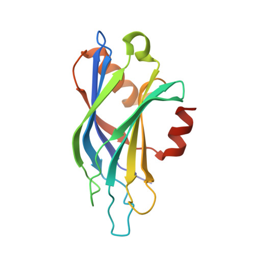
Architecture of the synaptotagmin-SNARE machinery for neuronal exocytosis.

Zhou, Q.Lai, Y.Bacaj, T.Zhao, M.Lyubimov, A.Y.Uervirojnangkoorn, M.Zeldin, O.B.Brewster, A.S.Sauter, N.K.Cohen, A.E.Soltis, S.M.Alonso-Mori, R.Chollet, M.Lemke, H.T.Pfuetzner, R.A.Choi, U.B.Weis, W.I.Diao, J.Sudhof, T.C.Brunger, A.T.

(2015) Nature 525: 62-67

  • DOI: https://doi.org/10.1038/nature14975
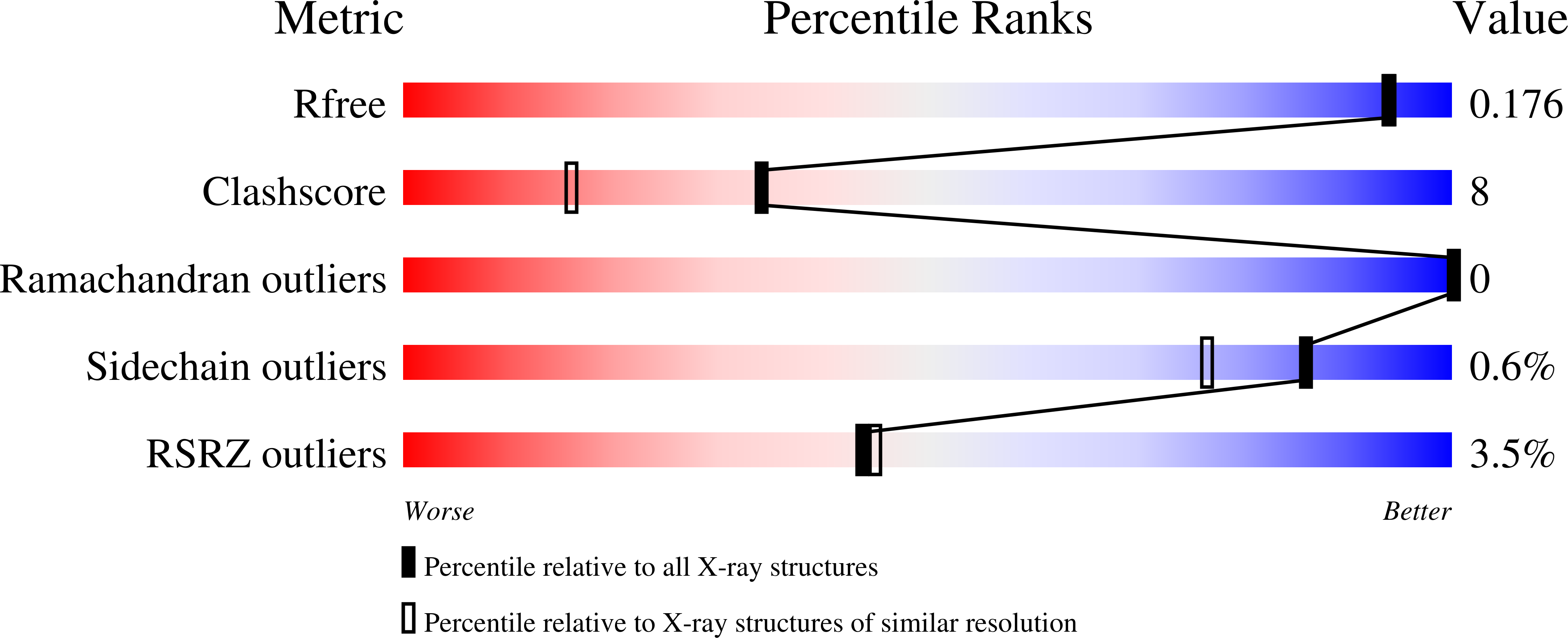
  • Primary Citation of Related Structures:  
    5CCG, 5CCH, 5CCI, 5CCJ

  • PubMed Abstract: 

    Synaptotagmin-1 and neuronal SNARE proteins have central roles in evoked synchronous neurotransmitter release; however, it is unknown how they cooperate to trigger synaptic vesicle fusion. Here we report atomic-resolution crystal structures of Ca(2+)- and Mg(2+)-bound complexes between synaptotagmin-1 and the neuronal SNARE complex, one of which was determined with diffraction data from an X-ray free-electron laser, leading to an atomic-resolution structure with accurate rotamer assignments for many side chains. The structures reveal several interfaces, including a large, specific, Ca(2+)-independent and conserved interface. Tests of this interface by mutagenesis suggest that it is essential for Ca(2+)-triggered neurotransmitter release in mouse hippocampal neuronal synapses and for Ca(2+)-triggered vesicle fusion in a reconstituted system. We propose that this interface forms before Ca(2+) triggering, moves en bloc as Ca(2+) influx promotes the interactions between synaptotagmin-1 and the plasma membrane, and consequently remodels the membrane to promote fusion, possibly in conjunction with other interfaces.


  • Organizational Affiliation

    Department of Molecular and Cellular Physiology, Howard Hughes Medical Institute, Stanford University, Stanford, California 94305, USA.


Macromolecules
Find similar proteins by:  (by identity cutoff)  |  3D Structure
Entity ID: 1
MoleculeChains Sequence LengthOrganismDetailsImage
Synaptotagmin-1
A, B, C, D
152Rattus norvegicusMutation(s): 5 
Gene Names: Syt1
UniProt
Find proteins for P21707 (Rattus norvegicus)
Explore P21707 
Go to UniProtKB:  P21707
Entity Groups  
Sequence Clusters30% Identity50% Identity70% Identity90% Identity95% Identity100% Identity
UniProt GroupP21707
Sequence Annotations
Expand
  • Reference Sequence
Small Molecules
Ligands 2 Unique
IDChains Name / Formula / InChI Key2D Diagram3D Interactions
SO4
Query on SO4

Download Ideal Coordinates CCD File 
AA [auth B]
AB [auth D]
BA [auth B]
CA [auth B]
DA [auth B]
AA [auth B],
AB [auth D],
BA [auth B],
CA [auth B],
DA [auth B],
K [auth A],
KA [auth C],
L [auth A],
LA [auth C],
M [auth A],
MA [auth C],
N [auth A],
O [auth A],
VA [auth D],
W [auth B],
WA [auth D],
X [auth B],
XA [auth D],
Y [auth B],
YA [auth D],
Z [auth B],
ZA [auth D]
SULFATE ION
O4 S
QAOWNCQODCNURD-UHFFFAOYSA-L
GOL
Query on GOL

Download Ideal Coordinates CCD File 
E [auth A]
EA [auth C]
F [auth A]
FA [auth C]
G [auth A]
E [auth A],
EA [auth C],
F [auth A],
FA [auth C],
G [auth A],
GA [auth C],
H [auth A],
HA [auth C],
I [auth A],
IA [auth C],
J [auth A],
JA [auth C],
NA [auth D],
OA [auth D],
P [auth B],
PA [auth D],
Q [auth B],
QA [auth D],
R [auth B],
RA [auth D],
S [auth B],
SA [auth D],
T [auth B],
TA [auth D],
U [auth B],
UA [auth D],
V [auth B]
GLYCEROL
C3 H8 O3
PEDCQBHIVMGVHV-UHFFFAOYSA-N
Experimental Data & Validation

Experimental Data

  • Method: X-RAY DIFFRACTION
  • Resolution: 1.65 Å
  • R-Value Free: 0.178 
  • R-Value Work: 0.150 
  • R-Value Observed: 0.151 
  • Space Group: P 21 21 21
Unit Cell:
Length ( Å )Angle ( ˚ )
a = 68.92α = 90
b = 107.59β = 90
c = 110.8γ = 90
Software Package:
Software NamePurpose
PHENIXrefinement
XSCALEdata scaling
PHASERphasing
PDB_EXTRACTdata extraction
XDSdata reduction

Structure Validation

View Full Validation Report


Entry History 

Deposition Data

Revision History  (Full details and data files)

  • Version 1.0: 2015-08-12
    Type: Initial release
  • Version 1.1: 2015-09-02
    Changes: Database references
  • Version 1.2: 2015-09-16
    Changes: Database references
  • Version 1.3: 2023-09-27
    Changes: Data collection, Database references, Derived calculations, Refinement description