Functional characterization of CYP107W1 from Streptomyces avermitilis and biosynthesis of macrolide oligomycin A.
Han, S., Pham, T.V., Kim, J.H., Lim, Y.R., Park, H.G., Cha, G.S., Yun, C.H., Chun, Y.J., Kang, L.W., Kim, D.(2015) Arch Biochem Biophys 575: 1-7
- PubMed: 25849761
- DOI: https://doi.org/10.1016/j.abb.2015.03.025
- Primary Citation of Related Structures:
4WPZ - PubMed Abstract:
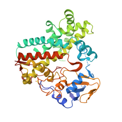
Streptomyces avermitilis contains 33 cytochrome P450 genes in its genome, many of which play important roles in the biosynthesis process of antimicrobial agents. Here, we characterized the biochemical function and structure of CYP107W1 from S. avermitilis, which is responsible for the 12-hydroxylation reaction of oligomycin C. CYP107W1 was expressed and purified from Escherichia coli. Purified proteins exhibited the typical CO-binding spectrum of P450. Interaction of oligomycin C and oligomycin A (12-hydroxylated oligomycin C) with purified CYP107W1 resulted in a type I binding with Kd values of 14.4 ± 0.7 μM and 2.0 ± 0.1 μM, respectively. LC-mass spectrometry analysis showed that CYP107W1 produced oligomycin A by regioselectively hydroxylating C12 of oligomycin C. Steady-state kinetic analysis yielded a kcat value of 0.2 min(-1) and a Km value of 18 μM. The crystal structure of CYP107W1 was determined at 2.1 Å resolution. The overall P450 folding conformations are well conserved, and the open access binding pocket for the large macrolide oligomycin C was observed above the distal side of heme. This study of CYP107W1 can help a better understanding of clinically important P450 enzymes as well as their optimization and engineering for synthesizing novel antibacterial agents and other pharmaceutically important compounds.
Organizational Affiliation:
Konkuk University, Department of Biological Sciences, Seoul 143-701, Republic of Korea.