4V3B

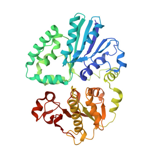
The structure of alpha2,3-sialyltransferase variant 1 from Pasteurella dagmatis in complex with the donor product CMP


Experimental Data Snapshot

  • Method: X-RAY DIFFRACTION
  • Resolution: 2.18 Å
  • R-Value Free: 0.238 
  • R-Value Work: 0.186 
  • R-Value Observed: 0.189 

wwPDB Validation   3D Report Full Report


Ligand Structure Quality Assessment 


This is version 1.1 of the entry. See complete history


Literature

Complete Switch from Alpha2,3- to Alpha2,6-Regioselectivity in Pasteurella Dagmatis Beta-D-Galactoside Sialyltransferase by Active-Site Redesign

Schmoelzer, K.Czabany, T.Pavkov-Keller, T.Luley-Goedl, C.Ribitsch, D.Schwab, H.Gruber, K.Nidetzky, B.

(2015) Chem Commun (Camb) 51: 3083

  • DOI: https://doi.org/10.1039/c4cc09772f

  • PubMed Abstract: 

    Structure-guided active-site redesign of a family GT-80 β-D-galactoside sialyltransferase (from Pasteurella dagmatis) to change enzyme regioselectivity from α-2,3 in the wild type to α-2,6 in a P7H-M117A double mutant is reported. Biochemical data for sialylation of lactose together with protein crystal structures demonstrate highly precise enzyme engineering.


  • Organizational Affiliation

    Austrian Centre of Industrial Biotechnology, Petersgasse 14, 8010 Graz, Austria.


Macromolecules
Find similar proteins by:  (by identity cutoff)  |  3D Structure
Entity ID: 1
MoleculeChains Sequence LengthOrganismDetailsImage
SIALYLTRANSFERASE388Pasteurella dagmatisMutation(s): 1 
UniProt
Find proteins for K9UUI6 (Pasteurella dagmatis)
Explore K9UUI6 
Go to UniProtKB:  K9UUI6
Entity Groups  
Sequence Clusters30% Identity50% Identity70% Identity90% Identity95% Identity100% Identity
UniProt GroupK9UUI6
Sequence Annotations
Expand
  • Reference Sequence
Small Molecules
Ligands 1 Unique
IDChains Name / Formula / InChI Key2D Diagram3D Interactions
C
Query on C

Download Ideal Coordinates CCD File 
B [auth A]CYTIDINE-5'-MONOPHOSPHATE
C9 H14 N3 O8 P
IERHLVCPSMICTF-XVFCMESISA-N
Experimental Data & Validation

Experimental Data

  • Method: X-RAY DIFFRACTION
  • Resolution: 2.18 Å
  • R-Value Free: 0.238 
  • R-Value Work: 0.186 
  • R-Value Observed: 0.189 
  • Space Group: P 65
Unit Cell:
Length ( Å )Angle ( ˚ )
a = 113.555α = 90
b = 113.555β = 90
c = 65.74γ = 120
Software Package:
Software NamePurpose
REFMACrefinement
XDSdata reduction
XDSdata scaling
PHASERphasing

Structure Validation

View Full Validation Report


Ligand Structure Quality Assessment 


Entry History 

Deposition Data

Revision History  (Full details and data files)

  • Version 1.0: 2015-04-08
    Type: Initial release
  • Version 1.1: 2024-01-10
    Changes: Data collection, Database references, Derived calculations, Other, Refinement description