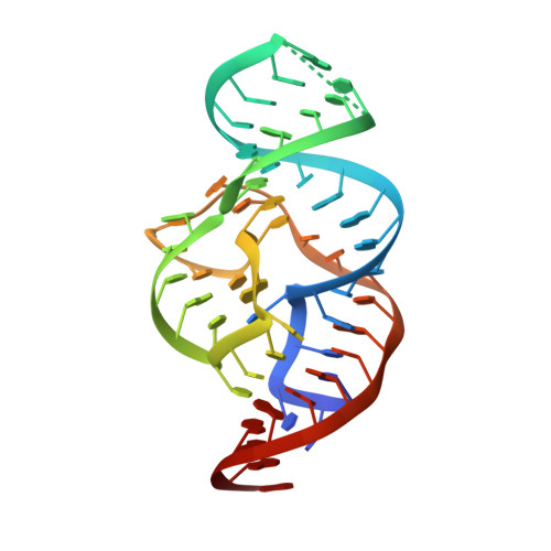
4RGF

Crystal structure of the in-line aligned env22 twister ribozyme soaked with Mn2+


Experimental Data Snapshot

  • Method: X-RAY DIFFRACTION
  • Resolution: 3.20 Å
  • R-Value Free: 0.277 
  • R-Value Work: 0.231 
  • R-Value Observed: 0.234 

wwPDB Validation   3D Report Full Report


This is version 1.1 of the entry. See complete history


Literature

In-line alignment and Mg(2+) coordination at the cleavage site of the env22 twister ribozyme.

Ren, A.Kosutic, M.Rajashankar, K.R.Frener, M.Santner, T.Westhof, E.Micura, R.Patel, D.J.

(2014) Nat Commun 5: 5534-5534

  • DOI: https://doi.org/10.1038/ncomms6534
  • Primary Citation of Related Structures:  
    4RGE, 4RGF

  • PubMed Abstract: 

    Small self-cleaving nucleolytic ribozymes contain catalytic domains that accelerate site-specific cleavage/ligation of phosphodiester backbones. We report on the 2.9-Å crystal structure of the env22 twister ribozyme, which adopts a compact tertiary fold stabilized by co-helical stacking, double-pseudoknot formation and long-range pairing interactions. The U-A cleavage site adopts a splayed-apart conformation with the modelled 2'-O of U positioned for in-line attack on the adjacent to-be-cleaved P-O5' bond. Both an invariant guanosine and a Mg(2+) are directly coordinated to the non-bridging phosphate oxygens at the U-A cleavage step, with the former positioned to contribute to catalysis and the latter to structural integrity. The impact of key mutations on cleavage activity identified an invariant guanosine that contributes to catalysis. Our structure of the in-line aligned env22 twister ribozyme is compared with two recently reported twister ribozymes structures, which adopt similar global folds, but differ in conformational features around the cleavage site.


  • Organizational Affiliation

    Structural Biology Program, Memorial Sloan-Kettering Cancer Center, New York, New York 10065, USA.


Macromolecules
Find similar nucleic acids by:  (by identity cutoff)  |  3D Structure
Entity ID: 1
MoleculeChains LengthOrganismImage
env22 twister ribozyme
A, B, C
56synthetic construct
Sequence Annotations
Expand
  • Reference Sequence
Small Molecules
Ligands 3 Unique
IDChains Name / Formula / InChI Key2D Diagram3D Interactions
MN
Query on MN

Download Ideal Coordinates CCD File 
AA [auth C]
D [auth A]
E [auth A]
F [auth A]
G [auth A]
AA [auth C],
D [auth A],
E [auth A],
F [auth A],
G [auth A],
H [auth A],
P [auth B],
Q [auth B],
X [auth C],
Y [auth C],
Z [auth C]
MANGANESE (II) ION
Mn
WAEMQWOKJMHJLA-UHFFFAOYSA-N
K
Query on K

Download Ideal Coordinates CCD File 
BA [auth C]
CA [auth C]
I [auth A]
J [auth A]
K [auth A]
BA [auth C],
CA [auth C],
I [auth A],
J [auth A],
K [auth A],
L [auth A],
M [auth A],
N [auth A],
O [auth A],
R [auth B],
S [auth B],
T [auth B],
U [auth B],
V [auth B]
POTASSIUM ION
K
NPYPAHLBTDXSSS-UHFFFAOYSA-N
MG
Query on MG

Download Ideal Coordinates CCD File 
W [auth C]MAGNESIUM ION
Mg
JLVVSXFLKOJNIY-UHFFFAOYSA-N
Experimental Data & Validation

Experimental Data

  • Method: X-RAY DIFFRACTION
  • Resolution: 3.20 Å
  • R-Value Free: 0.277 
  • R-Value Work: 0.231 
  • R-Value Observed: 0.234 
  • Space Group: P 61 2 2
Unit Cell:
Length ( Å )Angle ( ˚ )
a = 64.299α = 90
b = 64.299β = 90
c = 584.991γ = 120
Software Package:
Software NamePurpose
XDSdata scaling
PHENIXrefinement
XDSdata reduction

Structure Validation

View Full Validation Report


Entry History 

Deposition Data

Revision History  (Full details and data files)

  • Version 1.0: 2014-12-03
    Type: Initial release
  • Version 1.1: 2024-02-28
    Changes: Data collection, Database references, Derived calculations