


Crystal structure of Rv1600 encoded aminotransferase from Mycobacterium tuberculosis
Nasir, N., Anant, A., Vyas, R., Biswal, B.K.To be published.
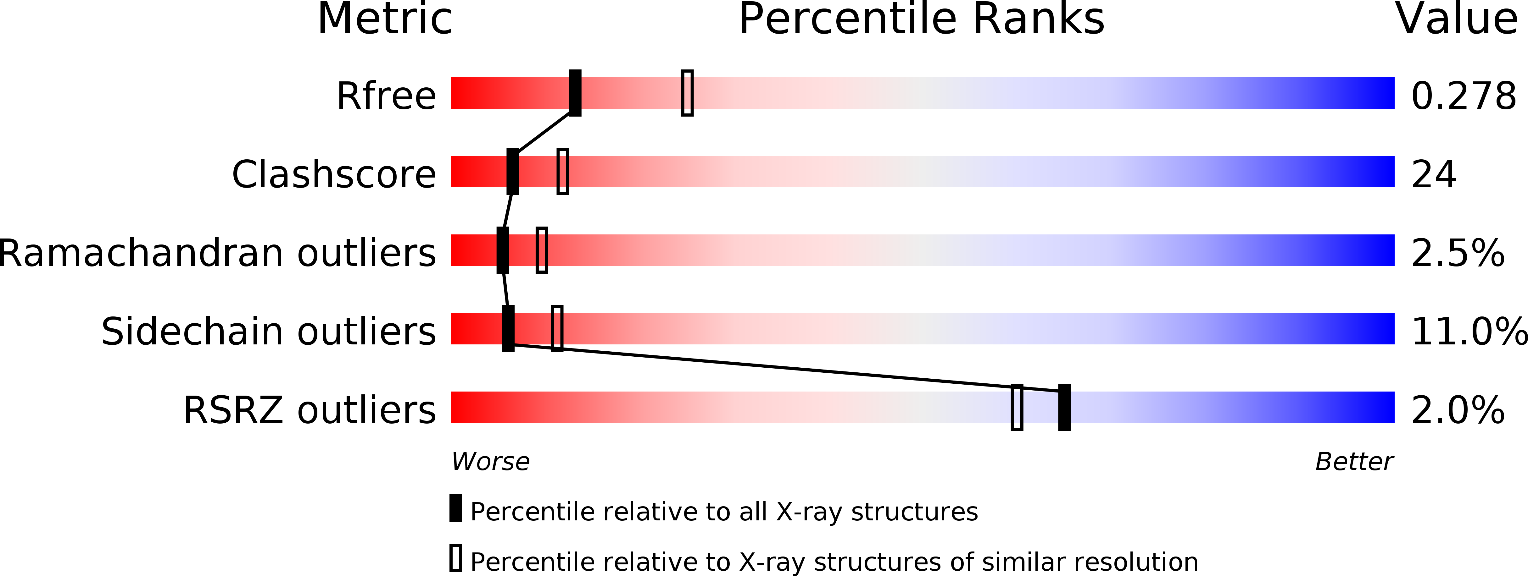
Experimental Data Snapshot
wwPDB Validation 3D Report Full Report
Entity ID: 1 | |||||
|---|---|---|---|---|---|
| Molecule | Chains | Sequence Length | Organism | Details | Image |
| Histidinol-phosphate aminotransferase | 394 | Mycobacterium tuberculosis H37Rv | Mutation(s): 0 Gene Names: hisC EC: 2.6.1.9 | 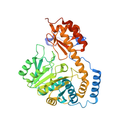 | |
UniProt | |||||
Find proteins for P9WML7 (Mycobacterium tuberculosis (strain ATCC 25618 / H37Rv)) Explore P9WML7 Go to UniProtKB: P9WML7 | |||||
Entity Groups | |||||
| Sequence Clusters | 30% Identity50% Identity70% Identity90% Identity95% Identity100% Identity | ||||
| UniProt Group | P9WML7 | ||||
Sequence AnnotationsExpand | |||||
| |||||
| Length ( Å ) | Angle ( ˚ ) |
|---|---|
| a = 159.937 | α = 90 |
| b = 159.937 | β = 90 |
| c = 110.317 | γ = 120 |
| Software Name | Purpose |
|---|---|
| StructureStudio | data collection |
| PHASER | phasing |
| REFMAC | refinement |
| HKL-2000 | data reduction |
| SCALEPACK | data scaling |
RCSB PDB (citation) is hosted by
RCSB PDB is a member of the

