


Design and Use of an Ester Analog of CoA to Trap the Michaelis Complex in a Thioesterase
Latham, J.A., Ji, T., Matthews, K., Allen, K.N., Dunaway-Mariano, D.To be published.
Experimental Data Snapshot
Entity ID: 1 | |||||
|---|---|---|---|---|---|
| Molecule | Chains | Sequence Length | Organism | Details | Image |
| Thioesterase PA1618 | 152 | Pseudomonas aeruginosa PAO1 | Mutation(s): 0 Gene Names: PA1618 EC: 3.1.2 | 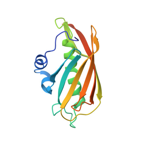 | |
UniProt | |||||
Find proteins for Q9I3A4 (Pseudomonas aeruginosa (strain ATCC 15692 / DSM 22644 / CIP 104116 / JCM 14847 / LMG 12228 / 1C / PRS 101 / PAO1)) Explore Q9I3A4 Go to UniProtKB: Q9I3A4 | |||||
Entity Groups | |||||
| Sequence Clusters | 30% Identity50% Identity70% Identity90% Identity95% Identity100% Identity | ||||
| UniProt Group | Q9I3A4 | ||||
Sequence AnnotationsExpand | |||||
| |||||
| Ligands 1 Unique | |||||
|---|---|---|---|---|---|
| ID | Chains | Name / Formula / InChI Key | 2D Diagram | 3D Interactions | |
| 0FQ Query on 0FQ | G [auth C], H [auth C], I [auth D], J [auth D] | phenacyl coenzyme A C29 H42 N7 O17 P3 S WOEFYXMDPBOUAP-VXAHOBLNSA-N |  | ||
| Length ( Å ) | Angle ( ˚ ) |
|---|---|
| a = 101.687 | α = 90 |
| b = 202.005 | β = 90 |
| c = 91.083 | γ = 90 |
| Software Name | Purpose |
|---|---|
| JBluIce-EPICS | data collection |
| PHENIX | model building |
| PHENIX | refinement |
| MOSFLM | data reduction |
| SCALA | data scaling |
| PHENIX | phasing |
RCSB PDB (citation) is hosted by
RCSB PDB is a member of the

