4OFP

Crystal Structure of SYG-2 D3-D4


Experimental Data Snapshot

  • Method: X-RAY DIFFRACTION
  • Resolution: 3.00 Å
  • R-Value Free: 0.283 
  • R-Value Work: 0.242 
  • R-Value Observed: 0.244 

wwPDB Validation   3D Report Full Report


Ligand Structure Quality Assessment 


This is version 1.2 of the entry. See complete history


Literature

Extracellular Architecture of the SYG-1/SYG-2 Adhesion Complex Instructs Synaptogenesis.

Ozkan, E.Chia, P.H.Wang, R.R.Goriatcheva, N.Borek, D.Otwinowski, Z.Walz, T.Shen, K.Garcia, K.C.

(2014) Cell 156: 482-494

  • DOI: https://doi.org/10.1016/j.cell.2014.01.004
  • Primary Citation of Related Structures:  
    4OF0, 4OF3, 4OF6, 4OF7, 4OF8, 4OFD, 4OFI, 4OFK, 4OFP, 4OFY

  • PubMed Abstract: 

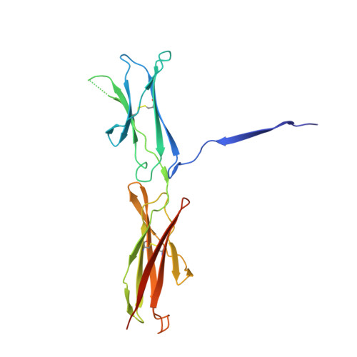
    SYG-1 and SYG-2 are multipurpose cell adhesion molecules (CAMs) that have evolved across all major animal taxa to participate in diverse physiological functions, ranging from synapse formation to formation of the kidney filtration barrier. In the crystal structures of several SYG-1 and SYG-2 orthologs and their complexes, we find that SYG-1 orthologs homodimerize through a common, bispecific interface that similarly mediates an unusual orthogonal docking geometry in the heterophilic SYG-1/SYG-2 complex. C. elegans SYG-1's specification of proper synapse formation in vivo closely correlates with the heterophilic complex affinity, which appears to be tuned for optimal function. Furthermore, replacement of the interacting domains of SYG-1 and SYG-2 with those from CAM complexes that assume alternative docking geometries or the introduction of segmental flexibility compromised synaptic function. These results suggest that SYG extracellular complexes do not simply act as "molecular velcro" and that their distinct structural features are important in instructing synaptogenesis. PAPERFLICK:


  • Organizational Affiliation

    Department of Molecular and Cellular Physiology and Department of Structural Biology, Stanford University School of Medicine, Stanford, CA 94305, USA; Howard Hughes Medical Institute, Stanford University School of Medicine, Stanford, CA 94305, USA.


Macromolecules
Find similar proteins by:  (by identity cutoff)  |  3D Structure
Entity ID: 1
MoleculeChains Sequence LengthOrganismDetailsImage
Protein SYG-2
A, B, C, D
208Caenorhabditis elegansMutation(s): 0 
Gene Names: C26G2.1CELE_C26G2.1syg-2
UniProt
Find proteins for Q9U3P2 (Caenorhabditis elegans)
Explore Q9U3P2 
Go to UniProtKB:  Q9U3P2
Entity Groups  
Sequence Clusters30% Identity50% Identity70% Identity90% Identity95% Identity100% Identity
UniProt GroupQ9U3P2
Sequence Annotations
Expand
  • Reference Sequence
Experimental Data & Validation

Experimental Data

  • Method: X-RAY DIFFRACTION
  • Resolution: 3.00 Å
  • R-Value Free: 0.283 
  • R-Value Work: 0.242 
  • R-Value Observed: 0.244 
  • Space Group: P 43 21 2
Unit Cell:
Length ( Å )Angle ( ˚ )
a = 121.24α = 90
b = 121.24β = 90
c = 178.16γ = 90
Software Package:
Software NamePurpose
Blu-Icedata collection
PHASERphasing
PHENIXrefinement
XDSdata reduction
XDSdata scaling

Structure Validation

View Full Validation Report


Ligand Structure Quality Assessment 


Entry History 

Deposition Data

Revision History  (Full details and data files)

  • Version 1.0: 2014-02-19
    Type: Initial release
  • Version 1.1: 2020-07-29
    Type: Remediation
    Reason: Carbohydrate remediation
    Changes: Data collection, Database references, Derived calculations, Structure summary
  • Version 1.2: 2023-09-20
    Changes: Data collection, Database references, Refinement description, Structure summary