4KCX

BRDT in complex with Dinaciclib


Experimental Data Snapshot

  • Method: X-RAY DIFFRACTION
  • Resolution: 2.00 Å
  • R-Value Free: 0.253 
  • R-Value Work: 0.209 
  • R-Value Observed: 0.211 

wwPDB Validation   3D Report Full Report


Ligand Structure Quality Assessment 


This is version 1.2 of the entry. See complete history


Literature

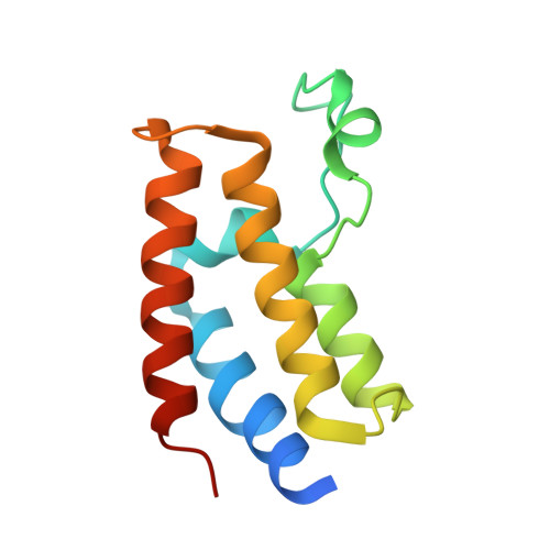
Cyclin-dependent kinase inhibitor dinaciclib interacts with the acetyl-lysine recognition site of bromodomains.

Martin, M.P.Olesen, S.H.Georg, G.I.Schonbrunn, E.

(2013) ACS Chem Biol 8: 2360-2365

  • DOI: https://doi.org/10.1021/cb4003283
  • Primary Citation of Related Structures:  
    4KCX, 4KD1

  • PubMed Abstract: 

    Bromodomain-containing proteins are considered atypical kinases, but their potential to interact with kinase inhibitors is unknown. Dinaciclib is a potent inhibitor of cyclin-dependent kinases (CDKs), which recently advanced to Phase III clinical trials for the treatment of leukemia. We determined the crystal structure of dinaciclib in complex with CDK2 at 1.7 Å resolution, revealing an elaborate network of binding interactions in the ATP site, which explains the extraordinary potency and selectivity of this inhibitor. Remarkably, dinaciclib also interacted with the acetyl-lysine recognition site of the bromodomain testis-specific protein BRDT, a member of the BET family of bromodomains. The binding mode of dinaciclib to BRDT at 2.0 Å resolution suggests that general kinase inhibitors ("hinge binders") possess a previously unrecognized potential to act as protein-protein inhibitors of bromodomains. The findings may provide a new structural framework for the design of next-generation bromodomain inhibitors using the vast chemical space of kinase inhibitors.


  • Organizational Affiliation

    Drug Discovery Department, H. Lee Moffitt Cancer Center and Research Institute , 12902 Magnolia Drive, Tampa, Florida 33612, United States.


Macromolecules
Find similar proteins by:  (by identity cutoff)  |  3D Structure
Entity ID: 1
MoleculeChains Sequence LengthOrganismDetailsImage
Bromodomain testis-specific protein
A, B
117Homo sapiensMutation(s): 0 
Gene Names: BRDT
UniProt & NIH Common Fund Data Resources
Find proteins for Q58F21 (Homo sapiens)
Explore Q58F21 
Go to UniProtKB:  Q58F21
PHAROS:  Q58F21
GTEx:  ENSG00000137948 
Entity Groups  
Sequence Clusters30% Identity50% Identity70% Identity90% Identity95% Identity100% Identity
UniProt GroupQ58F21
Sequence Annotations
Expand
  • Reference Sequence
Small Molecules
Ligands 2 Unique
IDChains Name / Formula / InChI Key2D Diagram3D Interactions
1QK
Query on 1QK

Download Ideal Coordinates CCD File 
C [auth A],
E [auth B]
3-[({3-ethyl-5-[(2S)-2-(2-hydroxyethyl)piperidin-1-yl]pyrazolo[1,5-a]pyrimidin-7-yl}amino)methyl]-1-hydroxypyridinium
C21 H29 N6 O2
WBUFFOKXERTKGU-SFHVURJKSA-N
CL
Query on CL

Download Ideal Coordinates CCD File 
D [auth A]CHLORIDE ION
Cl
VEXZGXHMUGYJMC-UHFFFAOYSA-M
Binding Affinity Annotations 
IDSourceBinding Affinity
1QK PDBBind:  4KCX Kd: 3.70e+4 (nM) from 1 assay(s)
Binding MOAD:  4KCX Kd: 3.70e+4 (nM) from 1 assay(s)
Experimental Data & Validation

Experimental Data

  • Method: X-RAY DIFFRACTION
  • Resolution: 2.00 Å
  • R-Value Free: 0.253 
  • R-Value Work: 0.209 
  • R-Value Observed: 0.211 
  • Space Group: P 1 21 1
Unit Cell:
Length ( Å )Angle ( ˚ )
a = 64.72α = 90
b = 29.95β = 94.12
c = 71.38γ = 90
Software Package:
Software NamePurpose
XDSdata scaling
PHASERphasing
PHENIXrefinement
XDSdata reduction
XSCALEdata scaling

Structure Validation

View Full Validation Report


Ligand Structure Quality Assessment 


Entry History 

Deposition Data

Revision History  (Full details and data files)

  • Version 1.0: 2013-09-18
    Type: Initial release
  • Version 1.1: 2013-12-25
    Changes: Database references
  • Version 1.2: 2023-09-20
    Changes: Data collection, Database references, Derived calculations, Refinement description